. 2014 Mar;42(5):3152-63.
doi: 10.1093/nar/gkt1280. Epub 2013 Dec 9.

Identification of residues required for stalled-ribosome rescue in the codon-independent release factor YaeJ

Affiliations

Identification of residues required for stalled-ribosome rescue in the codon-independent release factor YaeJ

Hiroyuki Kogure et al. Nucleic Acids Res. 2014 Mar.

Abstract

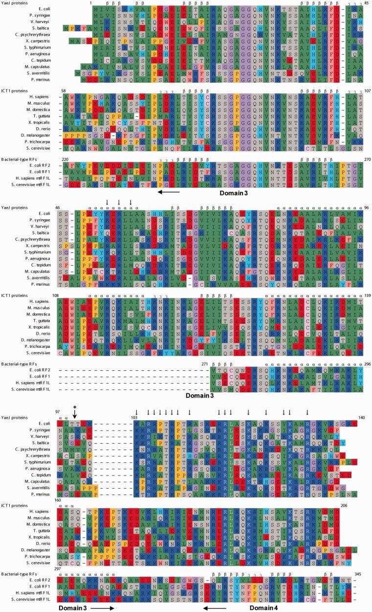
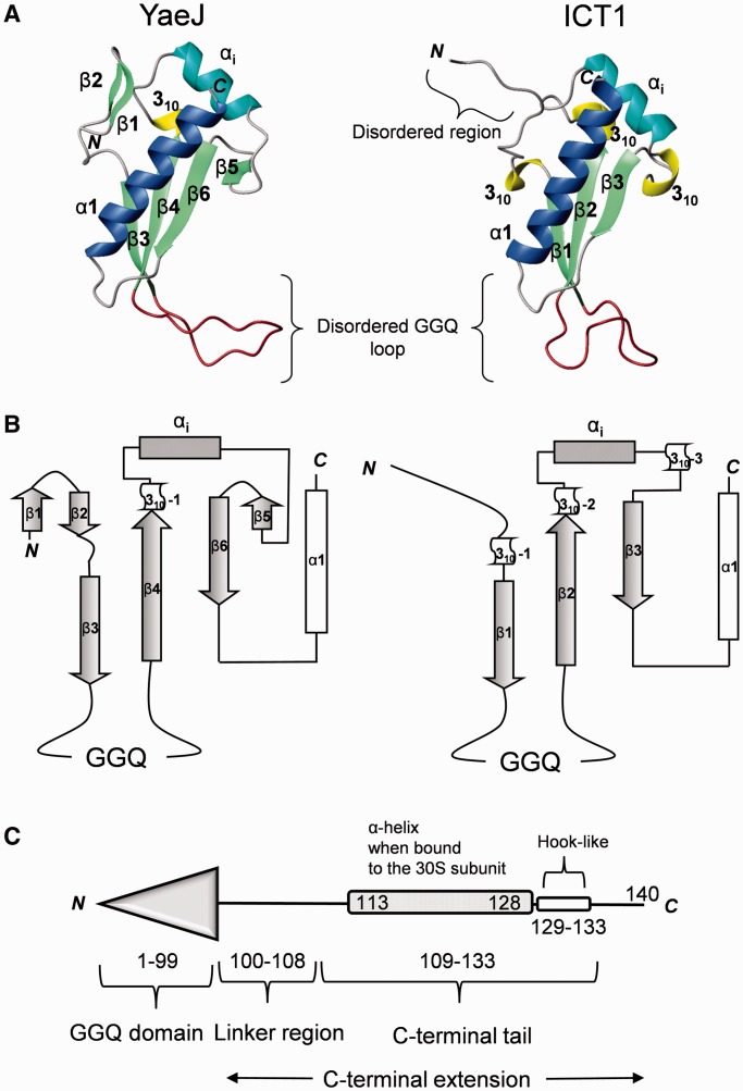
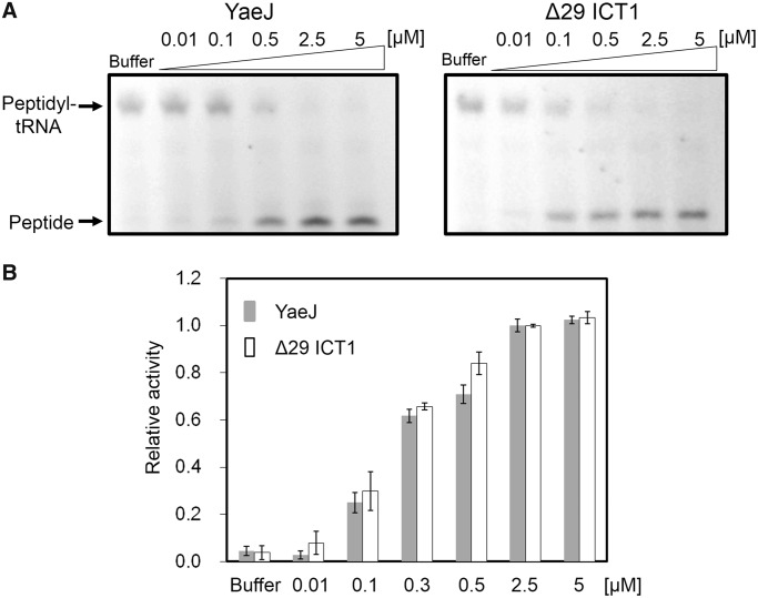
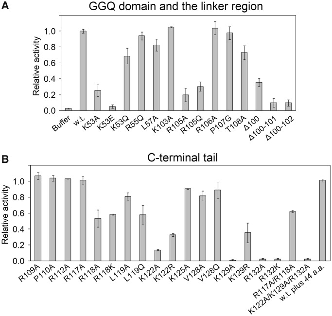
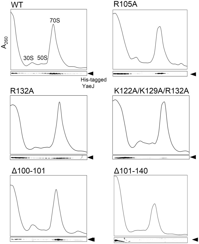
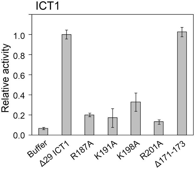
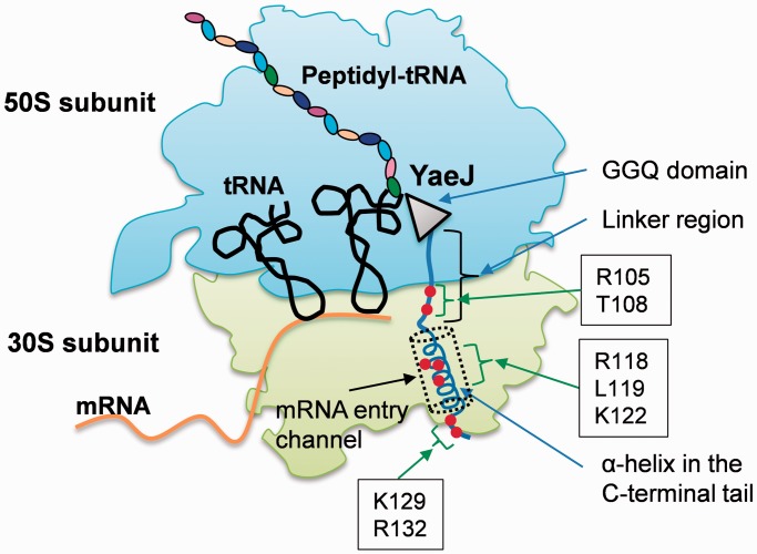
The YaeJ protein is a codon-independent release factor with peptidyl-tRNA hydrolysis (PTH) activity, and functions as a stalled-ribosome rescue factor in Escherichia coli. To identify residues required for YaeJ function, we performed mutational analysis for in vitro PTH activity towards rescue of ribosomes stalled on a non-stop mRNA, and for ribosome-binding efficiency. We focused on residues conserved among bacterial YaeJ proteins. Additionally, we determined the solution structure of the GGQ domain of YaeJ from E. coli using nuclear magnetic resonance spectroscopy. YaeJ and a human homolog, ICT1, had similar levels of PTH activity, despite various differences in sequence and structure. While no YaeJ-specific residues important for PTH activity occur in the structured GGQ domain, Arg118, Leu119, Lys122, Lys129 and Arg132 in the following C-terminal extension were required for PTH activity. All of these residues are completely conserved among bacteria. The equivalent residues were also found in the C-terminal extension of ICT1, allowing an appropriate sequence alignment between YaeJ and ICT1 proteins from various species. Single amino acid substitutions for each of these residues significantly decreased ribosome-binding efficiency. These biochemical findings provide clues to understanding how YaeJ enters the A-site of stalled ribosomes.


Figures

Figure 1.
Figure 1.
Sequence alignment of YaeJ proteins from bacteria, ICT1 proteins from eukaryotes and E. coli, and mitochondrial RFs. Secondary structure elements of the YaeJ structure determined in this study (PDB ID 2RTX), ICT1 (1J26) and the RF2 structure (1GQE) are indicated. The truncation position for the YaeJ protein used in the structural determination is marked by a vertical arrow with an asterisk. Vertical arrows indicate mutation positions, which are occupied by highly conserved residues (>90%) among all of the YaeJ proteins described in the article. Alignments are colored as follows: purple: glycine (G); yellow: proline (P); green: small and hydrophobic amino acids (A, V, L, I, M); pink: hydrophobic aromatic residues (F, W); gray: hydroxyl and amine amino acids (S, T, N, Q); red: negatively charged amino acids (D, E); blue: positively charged amino acids (K, R); pale pink: cysteine (C); cyan: histidine (H) and tyrosine (Y).
Figure 2.
Figure 2.
Comparison of the GGQ domains of YaeJ and ICT1. (A) Ribbon diagrams of the GGQ domain structures of E. coli YaeJ (PDB ID 2RTX; residues 1–109) and mouse ICT1 (PDB ID 1J26; residues 63–162). The α-helices α1 and αi are shown in blue and cyan, respectively. The 310 helices, β-strands, and the GGQ loop are shown in yellow, light green and brown, respectively. Note that the GGQ loops are disordered in both structures and thus their shapes vary. (B) Topology of their GGQ domain structures. (C) Schematic representations of YaeJ according to the co-crystal structure (PDB ID 4DH9).
Figure 3.
Figure 3.
YaeJ and ICT1 have similar PTH activity towards stalled ribosomes from E. coli. (A) In vitro translation of the non-stop template (non-stop) with recombinant His-tagged YaeJ (left) or ICT1 protein (Δ29 ICT1) (right). Each recombinant protein was added to the solution in which a 15-min in vitro translation reaction had been performed using the non-stop templates. The final concentration of YaeJ or Δ29 ICT1 is shown in each lane. (B) Extent of PTH given as the ratio of the band intensity of released peptides to that of peptidyl-tRNA plus that of released peptides at various protein concentrations. Released peptides indicate CRP proteins and peptidyl-tRNA indicates CRP-tRNA, respectively. Data are presented as mean ± SD for three independent experiments.
Figure 4.
Figure 4.
Effect of mutations of YaeJ on PTH activity. Wild-type YaeJ and the mutants in which well-conserved residues were substituted were expressed using the in vitro translation system. The in vitro translation reaction mixture containing YaeJ or a YaeJ mutant was directly added to another solution in which a preliminarily 15-min in vitro translation reaction had been performed using the non-stop template. The final concentration of YaeJ and the mutants was adjusted to 2.5 μM. Data are presented as mean ± SD for three independent experiments. (A) Mutations in the GGQ domain and the linker region. (B) Mutations in the C-terminal tail. The term ‘w.t. plus 44 a.a.’ means a mutant in which 44 residues derived from the expression plasmid pET15b were added to the C-terminus of the wild-type, as described previously (12).
Figure 5.
Figure 5.
Ribosome-binding properties of His-tagged recombinant YaeJ protein and the mutants. After separation through 5–20% sucrose gradients, fractions were analyzed by western blotting using an anti-His antibody. Results are representative of three independent experiments.
Figure 6.
Figure 6.
In vitro translation of the non-stop template (non-stop) with recombinant His-tagged ICT1 protein (Δ29 ICT1) and its mutants. The recombinant protein was added to the solution in which a 15-min in vitro translation reaction had been performed using the non-stop templates. Data are presented as mean ± SD from at least three independent experiments.
Figure 7.
Figure 7.
Mapping of residues required for PTH activity in YaeJ, as indicated by this study, onto a schematic representation of the YaeJ structure on a stalled ribosome. The schematic is based on the co-crystal structure of E. coli YaeJ and T. thermophilus 70S ribosome in complex with non-stop mRNA (PDB ID 4DH9). This study also showed that there is a minimum length for the linker region to achieve PTH activity. Note that the coordinates of residues 134–140 in the C-terminal extension are missing in the co-crystal structure.

References

    1. Moore SD, Sauer RT. The tmRNA system for translational surveillance and ribosome rescue. Annu. Rev. Biochem. 2007;76:101–124. - PubMed
    1. Janssen BD, Hayes CS. The tmRNA ribosome-rescue system. Adv. Protein Chem. Struct. Biol. 2012;86:151–191. - PMC - PubMed
    1. Keiler KC, Waller PR, Sauer RT. Role of a peptide tagging system in degradation of proteins synthesized from damaged messenger RNA. Science. 1996;271:990–993. - PubMed
    1. Himeno H, Sato M, Tadaki T, Fukushima M, Ushida C, Muto A. In vitro trans translation mediated by alanine-charged 10Sa RNA. J. Mol. Biol. 1997;268:803–808. - PubMed
    1. Karzai AW, Roche ED, Sauer RT. The SsrA-SmpB system for protein tagging, directed degradation and ribosome rescue. Nat. Struct. Biol. 2000;7:449–455. - PubMed

Publication types

MeSH terms

Associated data