Review
. 2014 May;71(10):1907-16.
doi: 10.1007/s00018-013-1535-6. Epub 2013 Dec 10.

Remodeling and dedifferentiation of adult cardiomyocytes during disease and regeneration

Affiliations
Review

Remodeling and dedifferentiation of adult cardiomyocytes during disease and regeneration

Marten Szibor et al. Cell Mol Life Sci. 2014 May.

Abstract

Cardiomyocytes continuously generate the contractile force to circulate blood through the body. Imbalances in contractile performance or energy supply cause adaptive responses of the heart resulting in adverse rearrangement of regular structures, which in turn might lead to heart failure. At the cellular level, cardiomyocyte remodeling includes (1) restructuring of the contractile apparatus; (2) rearrangement of the cytoskeleton; and (3) changes in energy metabolism. Dedifferentiation represents a key feature of cardiomyocyte remodeling. It is characterized by reciprocal changes in the expression pattern of "mature" and "immature" cardiomyocyte-specific genes. Dedifferentiation may enable cardiomyocytes to cope with hypoxic stress by disassembly of the energy demanding contractile machinery and by reduction of the cellular energy demand. Dedifferentiation during myocardial repair might provide cardiomyocytes with additional plasticity, enabling survival under hypoxic conditions and increasing the propensity to enter the cell cycle. Although dedifferentiation of cardiomyocytes has been described during tissue regeneration in zebrafish and newts, little is known about corresponding mechanisms and regulatory circuits in mammals. The recent finding that the cytokine oncostatin M (OSM) is pivotal for cardiomyocyte dedifferentiation and exerts strong protective effects during myocardial infarction highlights the role of cytokines as potent stimulators of cardiac remodeling. Here, we summarize the current knowledge about transient dedifferentiation of cardiomyocytes in the context of myocardial remodeling, and propose a model for the role of OSM in this process.


Conflict of interest statement

The authors declare that they have no conflicting commercial interests related to this work.

Figures
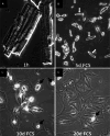
Fig. 1
Fig. 1
Morphology of cardiomyocytes in culture. Phase contrast micrographs of cultured adult rat cardiomyocytes freshly isolated (a) or after 5, 10 and 20 days in culture (b–d). Cultures were treated with 5 % fetal calf serum (FCS). White arrows cardiomyocytes round up apically and gradually loose cross-striated appearance. Black arrows rounded-up cardiomyocytes. Note the spreading of cardiomyocytes after FCS stimulation
Fig. 2
Fig. 2
Oncostatin M induces dedifferentiation of adult cardiomyocytes. Confocal images of adult rat cardiomyocytes after 7 days in culture stained with α-SM actin (red), sarcomeric α-actinin (green), and DAPI (blue). The upper panel represents merged images of all three channels (blue, red, green). Cultures were pretreated with 2 % FCS for 2 days and then kept in 2 % FCS (con) or stimulated with oncostatin M (OSM). Knock-down of the OSM receptor by siRNA (OSM + siOβ) prevents dedifferentiation. OSM treatment results in a massive loss of mature sarcomeres and morphological changes with typical cell extensions to re-establish cell-to-cell contacts. Scale bars left and middle rows 30 μm, right row 50 μm
Fig. 3
Fig. 3
Model of cardiomyocyte remodeling and dedifferentiation during cardiac regeneration and repair. Activation of oncostatin M (OSM) induces dedifferentiation and hibernation in surviving cardiomyocytes. Dedifferentiation might be reversed after revascularization and hypertrophic stimulation. Invading macrophages remove debris and release OSM in the damaged heart. Cardiomyocytes dedifferentiate in response to OSM and re-establish cell–cell contacts. Hypertrophic signals (IGF-1) induce re-differentiation and hypertrophy of cardiomyocytes. Extended presence of dedifferentiation signals compromises contractility and promotes adverse myocardial remodeling

Similar articles

Cited by

References

    1. Lopaschuk GD, Jaswal JS. Energy metabolic phenotype of the cardiomyocyte during development, differentiation, and postnatal maturation. J Cardiovasc Pharmacol. 2010;56(2):130–140. doi: 10.1097/FJC.0b013e3181e74a14. - DOI - PubMed
    1. Pohjoismaki JL, Boettger T, Liu Z, Goffart S, Szibor M, Braun T. Oxidative stress during mitochondrial biogenesis compromises mtDNA integrity in growing hearts and induces a global DNA repair response. Nucleic Acids Res. 2012;40(14):6595–6607. doi: 10.1093/nar/gks301. - DOI - PMC - PubMed
    1. Pohjoismaki JL, Kruger M, Al-Furoukh N, Lagerstedt A, Karhunen PJ, Braun T. Postnatal cardiomyocyte growth and mitochondrial reorganization cause multiple changes in the proteome of human cardiomyocytes. Mol BioSyst. 2013 - PubMed
    1. Rajabi M, Kassiotis C, Razeghi P, Taegtmeyer H. Return to the fetal gene program protects the stressed heart: a strong hypothesis. Heart Fail Rev. 2007;12(3–4):331–343. doi: 10.1007/s10741-007-9034-1. - DOI - PubMed
    1. Cohn JN, Ferrari R, Sharpe N. Cardiac remodeling–concepts and clinical implications: a consensus paper from an international forum on cardiac remodeling. Behalf of an international forum on cardiac remodeling. J Am Coll Cardiol. 2000;35(3):569–582. doi: 10.1016/S0735-1097(99)00630-0. - DOI - PubMed

Publication types

MeSH terms

LinkOut - more resources