. 2014 Jun;5(3):394-406.
doi: 10.1007/s12975-013-0304-z. Epub 2013 Nov 29.

Vascular neural network phenotypic transformation after traumatic injury: potential role in long-term sequelae

Affiliations

Vascular neural network phenotypic transformation after traumatic injury: potential role in long-term sequelae

J Badaut et al. Transl Stroke Res. 2014 Jun.

Abstract

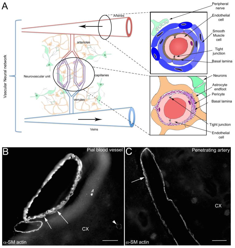
The classical neurovascular unit (NVU), composed primarily of endothelium, astrocytes, and neurons, could be expanded to include smooth muscle and perivascular nerves present in both the up- and downstream feeding blood vessels (arteries and veins). The extended NVU, which can be defined as the vascular neural network (VNN), may represent a new physiological unit to consider for therapeutic development in stroke, traumatic brain injury, and other brain disorders (Zhang et al., Nat Rev Neurol 8(12):711-716, 2012). This review is focused on traumatic brain injury and resultant post-traumatic changes in cerebral blood flow, smooth muscle cells, matrix, blood-brain barrier structures and function, and the association of these changes with cognitive outcomes as described in clinical and experimental reports. We suggest that studies characterizing TBI outcomes should increase their focus on changes to the VNN, as this may yield meaningful therapeutic targets to resolve posttraumatic dysfunction.


Conflict of interest statement

Conflict of Interest:

Jerome Badaut and Gregory Bix declare that they have no conflict of interest.

Figures

Figure 1
Figure 1
A) Schematic drawing of the vascular neural network (VNN, adapted from Zhang et al. [1]). The VNN includes the neurovascular unit (NVU), which is composed of endothelial cells, pericytes, basal lamina, astrocyte and neurons. The VNN also includes the cells that form and innervate larger blood vessels including endothelial cells, smooth muscle cells, and peripheral nerves. By definition, the feeding blood vessels running in the subarachnoid space are part of the VNN. This new physiological unit includes all the cells contributing to the maintenance of cerebral blood perfusion. B) Alpha Smooth muscle actin (α-SM actin) staining of pial blood vessels on the surface of the cortex (CX). A multilayer of SM cells is present (arrows) in the media of the pial blood vessel. Each of the individual SM cells may potentially have different functions, contractile and/or secretive. Staining of the contractile protein α-SM actin (arrowhead), present in pericytes, outlines some capillaries. C) α-SM actin staining in cortical penetrating blood vessel (CX) is outlining one or two layers of of smooth muscle cells (arrows) in the vascular wall of penetrating artery. In contrast with the vessels in the periphery and large cerebral feeding arteries, little is known about the effects of brain injury on phenotypic changes of the smooth muscle cells in the pial and penetrating blood vessels. Bar B, C = 40 μm
Figure 2
Figure 2
A, B) P-gp immunostaining (green) is present in endothelial cells, as shown in close proximity to the end-feet of GFAP-positive astrocytes (red) in both (A) sham and (C) juvenile TBI (jTBI). P-gp staining is significantly decreased in jTBI compared to sham animals at 2 months post-injury (kindly provided Pop et al. [115]). C, D) Caveolin-1 (Cav-1) staining is present in endothelial cells in sham (C) and jTBI (D) rats. Increased Cav-1 staining intensity in the jTBI compared to sham animals at 2 months post-injury reinforces the idea of phenotypic transformation of the endothelial cells at long-term post-TBI. Bar A, B = 50 μm; C, D= 100 μm
Figure 3
Figure 3
The schematic illustration describes the evolution of injury after TBI. The acute phase (blue box) after the primary injury has been largely studied for vascular dysfunction: with hypo-perfusion, hypo-metabolism and blood-brain barrier dysfunction. These changes have a direct consequence on neuronal cell death. The consequences of TBI on neuronal survival have been well documented. The question addressed here is: how do cerebral blood vessels recover in the long-term after traumatic brain injury? Clinical reports have demonstrated brain perfusion dysfunction at long-term after TBI (orange box). In jTBI, several changes in endothelial proteins (P-gp, Cav-1, claudin-5) have been observed up to two months post-TBI, suggesting a phenotypic transformation in the endothelium. Early vascular dysfunction (blue box) is still ongoing for several months/years after the primary injury and contributes to decreased blood flow, metabolism and pathological BBB. These cerebrovascular changes are possibly contributing to the absence of recovery in cognitive functions and some neurodegenerative processes observed in TBI patients.

References

    1. Zhang JH, Badaut J, Tang J, Obenaus A, Hartman R, Pearce WJ. The vascular neural network-a new paradigm in stroke pathophysiology. Nat Rev Neurol. 2012;8(12):711–6. doi: 10.1038/nrneurol.2012.210. - DOI - PMC - PubMed
    1. Coronado VG, Xu L, Basavaraju SV, McGuire LC, Wald MM, Faul MD, et al. Surveillance for traumatic brain injury-related deaths--United States, 1997–2007. MMWR Surveill Summ. 2011;60(5):1–32. ss6005a1 [pii] - PubMed
    1. Thurman D, Guerrero J. Trends in hospitalization associated with traumatic brain injury. JAMA. 1999;282(10):954–7. joc91173 [pii] - PubMed
    1. Pop V, Badaut J. A Neurovascular Perspective for Long-Term Changes After Brain Trauma. Translational stroke research. 2011;2(4):533–45. doi: 10.1007/s12975-011-0126-9. - DOI - PMC - PubMed
    1. Smith DH, Uryu K, Saatman KE, Trojanowski JQ, McIntosh TK. Protein accumulation in traumatic brain injury. Neuromolecular Med. 2003;4(1–2):59–72. doi: 10.1385/NMM:4:1-2:59. NMM:4:1-2:59 [pii] - DOI - PubMed

Publication types

LinkOut - more resources