. 2014 Mar;63(3):595-606.
doi: 10.1161/HYPERTENSIONAHA.113.02458. Epub 2013 Dec 9.

Excess LIGHT contributes to placental impairment, increased secretion of vasoactive factors, hypertension, and proteinuria in preeclampsia

Affiliations

Excess LIGHT contributes to placental impairment, increased secretion of vasoactive factors, hypertension, and proteinuria in preeclampsia

Wei Wang et al. Hypertension. 2014 Mar.

Abstract

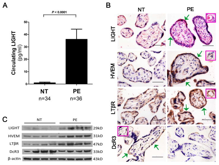
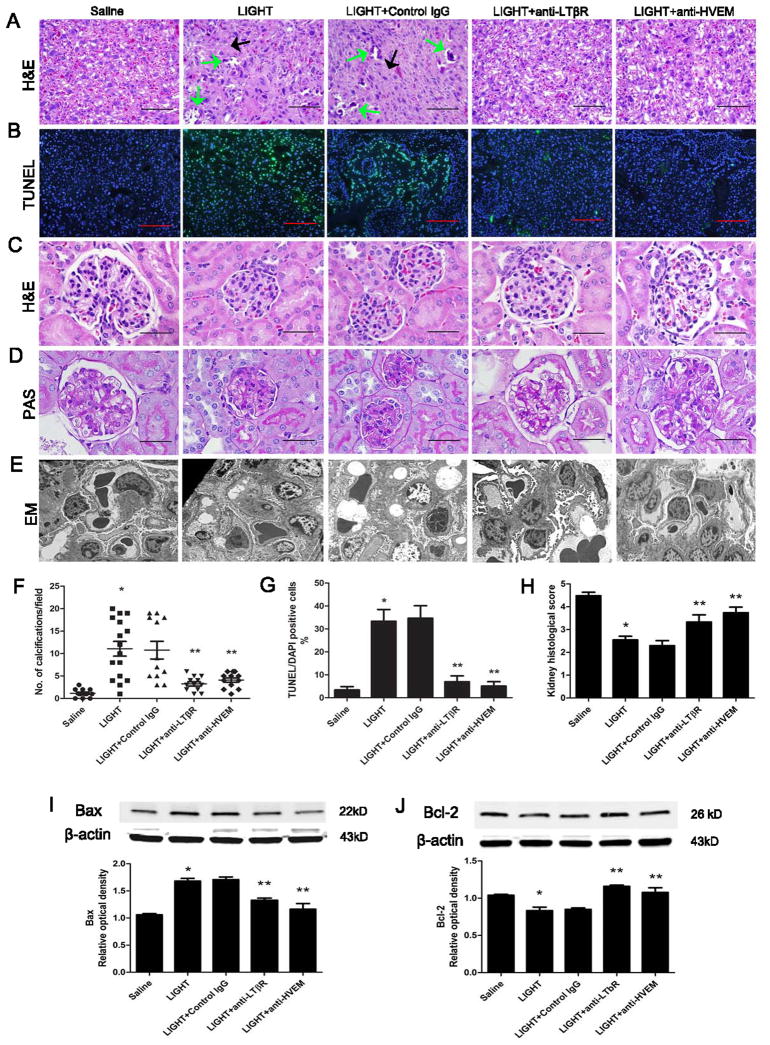
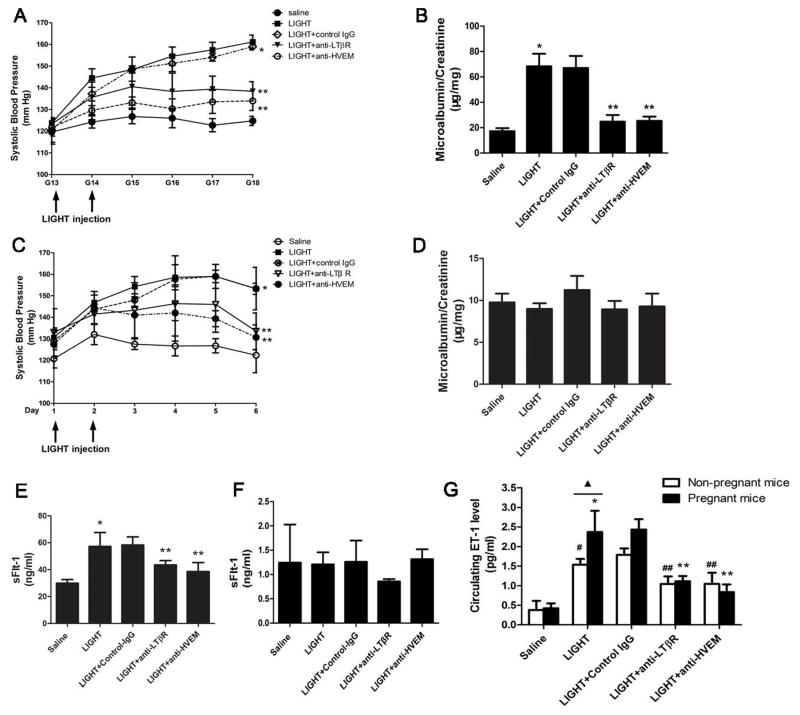
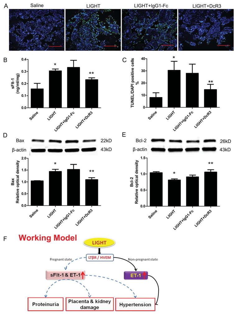
Preeclampsia, a prevalent hypertensive disorder of pregnancy, is believed to be secondary to uteroplacental ischemia. Accumulating evidence indicates that hypoxia-independent mediators, including inflammatory cytokines and growth factors, are associated with preeclampsia, but it is unclear whether these signals directly contribute to placental damage and disease development in vivo. We report that LIGHT, a novel tumor necrosis factor superfamily member, is significantly elevated in the circulation and placentas of preeclamptic women compared with normotensive pregnant women. Injection of LIGHT into pregnant mice induced placental apoptosis, small fetuses, and key features of preeclampsia, hypertension and proteinuria. Mechanistically, using neutralizing antibodies specific for LIGHT receptors, we found that LIGHT receptors herpes virus entry mediator and lymphotoxin β receptor are required for LIGHT-induced placental impairment, small fetuses, and preeclampsia features in pregnant mice. Accordingly, we further revealed that LIGHT functions through these 2 receptors to induce secretion of soluble fms-like tyrosine kinase-1 and endothelin-1, 2 well-accepted pathogenic factors in preeclampsia, and thereby plays an important role in hypertension and proteinuria in pregnant mice. Lastly, we extended our animal findings to human studies and demonstrated that activation of LIGHT receptors resulted in increased apoptosis and elevation of soluble fms-like tyrosine kinase-1 secretion in human placental villous explants. Overall, our human and mouse studies show that LIGHT signaling is a previously unrecognized pathway responsible for placental apoptosis, elevated secretion of vasoactive factors, and subsequent maternal features of preeclampsia, and reveal new therapeutic opportunities for the management of the disease.

Keywords: endothelin-1; herpes virus entry mediator; lymphotoxin-beta receptor; preeclampsia; receptors, tumor necrosis factor, member 14; tumor necrosis factor ligand superfamily member 14.


Figures

Figure 1
Figure 1. Circulating levels of LIGHT and placental expression profiles of LIGHT and its receptors in women with normotensive and PE pregnancies
(A) LIGHT levels were significantly increased in the circulation of preeclamptic patients (n=36) compared with normotensive pregnant women (n=34, P < 0.0001). (B) Localization of LIGHT and its receptors in the placentas was determined by immunohistochemistry. LIGHT and three receptors LTβR, HVEM and DcR3 were all present in human placental tissue-trophoblast cells (green arrows) and endothelial cells (inset box); (Scale bar = 100μm). (C) Western blotting revealed that protein levels of LIGHT and its transmembrane receptors LTβR and HVEM were elevated in placental tissue from preeclamptic patients (n=4) compared to normotensive pregnant women (n=4). However, the protein level of decoy receptor DcR3 was decreased in the placental tissue of pleeclamptic patients.
Figure 2
Figure 2. LIGHT-induced placental damage, apoptosis and kidney injury in pregnant mice by activation of LTβR and HVEM
(A) Placentas assessed by H&E staining indicated that LIGHT-injected pregnant mice had damaged placentas: calcifications (green arrows) and fibrotic areas (black arrows). Co-injection of neutralizing antibodies to LTβR or HVEM significantly attenuated placental damage (scale bar=100μm). (B) Placental apoptosis was assessed by TUNEL staining (×10 magnification; green, TUNEL-positive cells; blue, DAPI nuclear stain; scale bar: 1 mm). LIGHT injected pregnant mice had increased apoptosis in the labyrinth zones of their placentas as compared with saline-injected animals. (C–E) Kidneys assessed by H&E staining (C), PAS staining (D) and EM studies (E) indicated that LIGHT-injected pregnant mice had damaged kidneys with typical endotheliosis with decreased capillary lumens with swollen endothelial cells. Co-injection of neutralizing antibodies to LTβR or HVEM significantly attenuated kidney injury (scale bar=100μm). (F) An arbitrary histological quantification of the number of calcifications obtained per field under ×10 magnification (12 placentas for each group). (G) Quantification of the TUNEL assay indicated elevation of the TUNEL-positive cells in the placenta of the mice injected with LIGHT. Co-injection of neutralizing antibodies for LTβR or HVEM reduced the TUNEL-positive cells in the placentas as compared with the LIGHT injected animals (n=10 placentas per variable, from 5 different mice in each group). (H) Kidney histological score in pregnant mice was significantly increased with LIGHT injection. Co-injection of neutralizing antibodies to LTβR or HVEM significantly reduced the kidney histological score. (I–J) Western blot analysis of mouse placentas indicated that LIGHT injection led to increased pro-apoptotic protein Bax (I) and decreased anti-apoptotic protein Bcl-2 (J); n=5 for each variable collected over four independent experiments. Co-injection of neutralizing antibodies for LTβR or HVEM significantly reduced LIGHT-induced apoptotic features. Data are expressed as means ± SEM. *P < 0.05 versus saline injected pregnant mice; ** P < 0.05 versus LIGHT injected pregnant mice.
Figure 3
Figure 3. LIGHT signaling through LTβR and HVEM induced hypertension and proteinuria in the pregnant mice by increasing both sFlt-1 and ET-1 secretion. However, LIGHT only induced hypertension but not proteinuria in non-pregnant mice by increasing secretion of ET-1 and not sFlt-1
(A–B) Pregnant mice were injected with saline or LIGHT with or without injection of neutralizing antibodies for LTβR and HVEM, respectively. LIGHT injection resulted in hypertension (A) and proteinuria (B) in pregnant mice. Neutralizing antibodies for LTβR or HVEM significantly reduced hypertension seen in these mice (* P<0.001 versus saline injected mice; ** P<0.01 versus LIGHT injected mice). (C–D) LIGHT induced only hypertension (C) but not proteinuria (D) in non-pregnant mice. Neutralizing antibodies for LTβR or HVEM significantly attenuated hypertension in these mice (n=5–7). (* P<0.01 versus saline injected mice; ** P<0.05 versus LIGHT injected mice). (E–F) Circulating sFlt-1 level was significantly increased in LIGHT-injected pregnant mice (E) but not in non-pregnant mice (F). Neutralizing antibodies for LTβR and HVEM significantly attenuated LIGHT-induced sFlt-1 production in pregnant mice (E) but had no effect on the low sFlt-1 levels in non-pregnant mice (F). (G) Circulating ET-1 levels were remarkably elevated in LIGHT-injected pregnant and non-pregnant mice; and the LIGHT-mediated increase of circulating ET-1 in pregnant mice was significantly higher than that of non-pregnant mice (P<0.05). Neutralizing antibodies for LTβR or HVEM significantly attenuated LIGHT induced ET-1 production in pregnant mice and non-pregnant mice (*P<0.05 versus saline injected pregnant mice; ** P < 0.05 versus to LIGHT injected pregnant mice; #P<0.05 versus saline injected non-pregnant mice; ##P<0.05 versus LIGHT injected non-pregnant mice). Control rat IgG had no effect on LIGHT induced hypertension, proteinuria or sFlt-1 and ET-1 production.
Figure 4
Figure 4. LIGHT–mediated increase in placental tissue apoptosis and sFlt-1 secretion from human placental villous explants are inhibited by blocking LTβR or HVEM
Human placental villous explants treated with saline or LIGHT in the presence or absence of DcR3 or IgG-Fc. (A) TUNEL-stained cultured human villous explants indicated that LIGHT increased apoptosis (green, TUNEL-positive cells; blue, DAPI nuclear stain; Bars, 500 μm). (B) sFlt-1 secretion from cultured human villous explants was significantly increased after 24 hour-incubation with LIGHT compared to saline-treated human villous explants. DcR3 significantly reduced sFlt-1 secretion induced by LIGHT (n=6 for each group). (C) Quantification of the increased apoptosis is reflected in an increased apoptotic index in the explants incubated with LIGHT. Co-incubation of the explants with LIGHT and DcR3 partially attenuated the increase in cell death. (D–E) Western blot analysis of explant proteins demonstrate increased Bax (D) and decreased Bcl-2 (E), indicating a proapoptotic state. Explants of placentas from two different women were cultured, and each variable was examined six times per placenta (n=12). IgG1-Fc had no effect on LIGHT-induced placental apoptosis and sFlt-1 secretion from human placental villous explants. Data are expressed as means ± SEM. *P < 0.05 compared to saline incubated group; **P<0.05 compared to LIGHT-incubated group. (F) Working Model: Circulating LIGHT level is increased in PE patients. Elevated LIGHT functioning via activation of two transmembrane receptors, LTβR and HVEM, contributes to placental damage featured with apoptosis, secretion of vasoactive factors including sFlt-1 and ET-1 and subsequent disease development including hypertension and proteinuria in pregnant mice. However, LIGHT signaling via LTβR and HVEM only induces hypertension, but not proteinuria, in non-pregnant mice. LIGHT-mediated sFlt-1 production is a key mechanism underlying placenta tissue damage and preeclampsia development. Elevated of ET-1 is a common factor mediating LIGHT induced hypertension in both pregnant mice and non-pregnant mice. Decoy receptor, DcR3, can attenuate sFlt-1 secretion and placenta apoptosis stimulated by LIGHT. Thus, LIGHT signaling is a novel pathway contributing to pathogenesis of PE and serves as a potential therapeutic target for the disease.

Similar articles

Cited by

References

    1. Roberts JM, Cooper DW. Pathogenesis and genetics of pre-eclampsia. Lancet. 2001;357:53–56. - PubMed
    1. Granger JP, Alexander BT, Llinas MT, Bennett WA, Khalil RA. Pathophysiology of preeclampsia: Linking placental ischemia/hypoxia with microvascular dysfunction. Microcirculation. 2002;9:147–160. - PubMed
    1. Godfrey KM, Barker DJ. Fetal nutrition and adult disease. The American journal of clinical nutrition. 2000;71:1344S–1352S. - PubMed
    1. Barker DJ. In utero programming of chronic disease. Clin Sci (Lond) 1998;95:115–128. - PubMed
    1. Saito S, Sakai M. Th1/th2 balance in preeclampsia. Journal of reproductive immunology. 2003;59:161–173. - PubMed

Publication types

Substances