. 2013 Dec 24;110(52):21095-100.
doi: 10.1073/pnas.1319784110. Epub 2013 Dec 9.

The adjuvant MF59 induces ATP release from muscle that potentiates response to vaccination

Affiliations

The adjuvant MF59 induces ATP release from muscle that potentiates response to vaccination

Maria Vono et al. Proc Natl Acad Sci U S A. .

Abstract

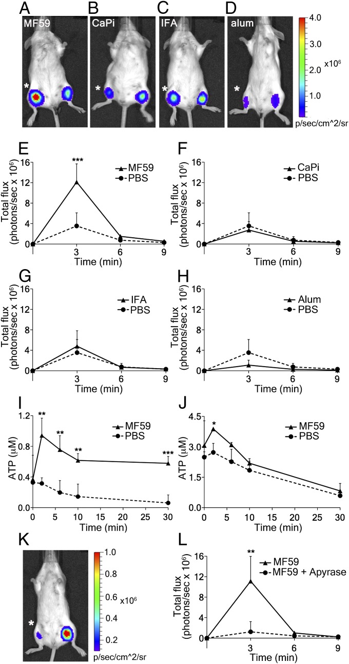
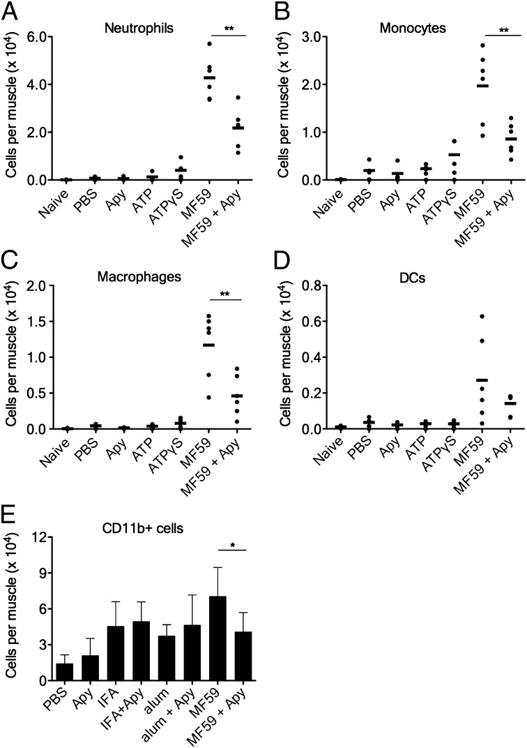
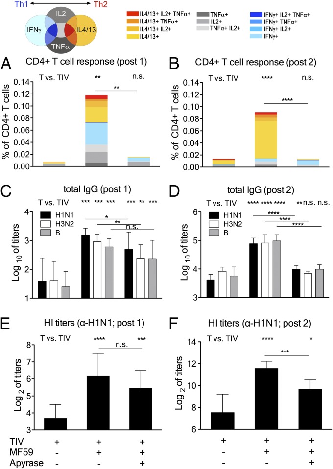
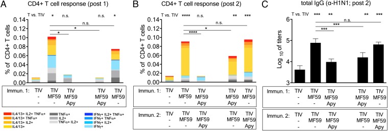
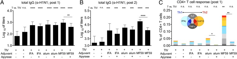
Vaccines are the most effective agents to control infections. In addition to the pathogen antigens, vaccines contain adjuvants that are used to enhance protective immune responses. However, the molecular mechanism of action of most adjuvants is ill-known, and a better understanding of adjuvanticity is needed to develop improved adjuvants based on molecular targets that further enhance vaccine efficacy. This is particularly important for tuberculosis, malaria, AIDS, and other diseases for which protective vaccines do not exist. Release of endogenous danger signals has been linked to adjuvanticity; however, the role of extracellular ATP during vaccination has never been explored. Here, we tested whether ATP release is involved in the immune boosting effect of four common adjuvants: aluminum hydroxide, calcium phosphate, incomplete Freund's adjuvant, and the oil-in-water emulsion MF59. We found that intramuscular injection is always associated with a weak transient release of ATP, which was greatly enhanced by the presence of MF59 but not by all other adjuvants tested. Local injection of apyrase, an ATP-hydrolyzing enzyme, inhibited cell recruitment in the muscle induced by MF59 but not by alum or incomplete Freund's adjuvant. In addition, apyrase strongly inhibited influenza-specific T-cell responses and hemagglutination inhibition titers in response to an MF59-adjuvanted trivalent influenza vaccine. These data demonstrate that a transient ATP release is required for innate and adaptive immune responses induced by MF59 and link extracellular ATP with an enhanced response to vaccination.

Keywords: DAMP; danger associated molecular pattern; inflammation; vaccine adjuvants.


Conflict of interest statement

The authors declare no conflict of interest.

Figures

Fig. 1.
Fig. 1.
Adjuvant-induced ATP release in mouse muscles and the effect of apyrase. (AD) Representative images taken 3 min after intramuscular injection of adjuvants (right hind limb; asterisk) or PBS (left hind limb) in BALB/c mice together with the mixture luciferase-luciferin that reports on ATP changes. (EH) Corresponding quantitative analyses of chemiluminescence emission over time (number of photons per second in the region of interest). (A and E) MF59 (40% vol/vol), (B and F) CaPi (50 μg), (C and G) IFA (40% vol/vol), (D and H) alum (100 μg), and (I and J) ATP release from ex vivo mouse muscles injected with MF59. Mouse tibialis anterior (I) and quadriceps (J) muscles were exposed and injected with MF59 (40% vol/vol), continuous lines; the dotted line refers to the injection of the same volume of PBS. Muscles were rapidly removed and suspended in oxygenated buffer at 37 °C. ATP released into buffer was quantified at the given times, using the luciferin-luciferase assay and a known ATP standard. (K) Representative image taken 3 min after intramuscular injection of MF59 + apyrase (10 U; asterisk) or MF59 alone (contralateral muscle). (L) Corresponding quantitative analysis over time. Data show mean values + SD from at least four independent experiments. Unpaired, two-tailed Student’s t test (T): *P < 0.05, **P < 0.01, ***P < 0.001.
Fig. 2.
Fig. 2.
Coinjection of apyrase reduces immune cell recruitment induced by MF59, but not by IFA or alum. (AD) Groups of mice were injected i.m. with the indicated compounds at the following doses: MF59 (40% vol/vol), ATP or ATP-γS (5mM), apyrase (Apy) (10 U per leg), or PBS. Single-cell suspensions of treated muscles were analyzed by FACS 24 h postinjection. Dots show numbers of the respective cell type per individual muscle (N ≥ 4 per group), whereas black bars indicate arithmetic means. (A) Neutrophils, (B) monocytes, (C) macrophages, and (D) dendritic cells (DCs). (E) Groups of mice were injected i.m. with the indicated compounds at the following doses: MF59 (20% vol/vol), IFA (40% vol/vol), alum (100 μg), apyrase (10 U per leg), or PBS, all in presence of OVA (10 μg per mouse). Numbers of CD11b+ cells are reported, data show mean values + SD from eight to 12 muscles per group. Unpaired, two-tailed Student’s t test (T): *P < 0.05, **P < 0.01.
Fig. 3.
Fig. 3.
Coinjection of apyrase inhibits adjuvanticity of MF59 to a trivalent influenza vaccine (TIV). (AF) twelve mice per group were immunized twice (4 wk apart) with TIV and adjuvants, as indicated: MF59 (40% vol/vol), apyrase (10 U per leg), and TIV (0.1 μg each antigen). (A and B) Spleens from 4 mice per group were taken 2 wk after each immunization, and vaccine-specific CD4+ T helper cells were reactivated by in vitro stimulation. Their individual cytokine profile was assessed by intracellular cytokine staining and FACS analysis. The bars show cumulative numbers of vaccine-specific cytokine expressing cells after the first (A) and second (B) immunization, and the individual color code indicates the type of cytokines expressed by the respective cells, as indicated. (CF) Serum samples were drawn 2 wk after each immunization, and vaccine-specific antibody titers were measured. Total IgG antibody titers toward H1N1/California, H3N2/Perth, and B/Brisbane after the first (C) and second (D) immunization. Values represent mean logarithmic titers (log 10) of eight to 12 mice per group + SD. Hemagglutination inhibition titers toward H1N1/California after the first (E) and second (F) immunization; values represent means of Log2 titers of eight to 12 mice per group + SD. Unpaired, two-tailed Student’s t test (T): *P < 0.05,**P < 0.01, ***P < 0.001,****P < 0.0001.
Fig. 4.
Fig. 4.
ATP release induced by MF59 is essential during the first vaccination. Mice were immunized as before with TIV and adjuvants, as indicated. One group of mice received two doses of MF59+apyrase, whereas in the other groups, apyrase (Apy) was added to MF59 only during prime or only during boost. (A and B) Spleens from 4 mice per group were taken 2 wk after each immunization, and vaccine-specific CD4+ T helper cells were reactivated by in vitro stimulation, as before. The bars show cumulative numbers of vaccine-specific cytokine expressing cells after the first (A) and second (B) immunization, whereas the individual color code indicates the type of cytokines expressed by the respective cells. (C) Serum samples were drawn 2 wk after the second immunization, and total IgG antibody titers toward H1N1/California were measured by ELISA. Values represent mean logarithmic titers (log 10) of eight mice per group + SD. Unpaired, two-tailed Student’s t test (T): *P < 0.05, **P < 0.01, ***P < 0.001, ****P < 0.0001.
Fig. 5.
Fig. 5.
Apyrase inhibits antibody responses induced by TIV adjuvanted with MF59, but not with alum and IFA. (AC) Twelve mice per group were immunized as before with TIV and adjuvants, as indicated. The following doses were used: MF59 (40% vol/vol), IFA (40% vol/vol), alum (100 μg), apyrase (10 U per leg), and TIV (0.1 μg each antigen). (A and B) Serum samples were drawn 2 wk after the first (A) or second (B) immunization, and total IgG antibody titers toward H1N1/California were measured by ELISA. Values represent mean logarithmic titers (log 10) of eight to 12 mice per group + SD. (C) Spleens from 4 mice per group were taken 2 wk after the first immunization, and vaccine-specific CD4+ T helper cells were reactivated by in vitro stimulation. Their individual cytokine profile was assessed by intracellular cytokine staining and FACS analysis. The bars show cumulative numbers of vaccine-specific cytokine expressing cells, as indicated. Unpaired, two-tailed Student’s t test (T): *P < 0.05, **P < 0.01, ***P < 0.001, ****P < 0.0001.

References

    1. McKee AS, MacLeod MKL, Kappler JW, Marrack P. Immune mechanisms of protection: Can adjuvants rise to the challenge? BMC Biol. 2010;8:37. - PMC - PubMed
    1. McKee AS, Munks MW, Marrack P. How do adjuvants work? Important considerations for new generation adjuvants. Immunity. 2007;27(5):687–690. - PubMed
    1. Coffman RL, Sher A, Seder RA. Vaccine adjuvants: Putting innate immunity to work. Immunity. 2010;33(4):492–503. - PMC - PubMed
    1. Lambrecht BN, Kool M, Willart MA, Hammad H. Mechanism of action of clinically approved adjuvants. Curr Opin Immunol. 2009;21(1):23–29. - PubMed
    1. Tritto E, Mosca F, De Gregorio E. Mechanism of action of licensed vaccine adjuvants. Vaccine. 2009;27(25-26):3331–3334. - PubMed

Publication types

MeSH terms