. 2014 Jan;3(1):18-31.
doi: 10.5966/sctm.2013-0110. Epub 2013 Dec 9.

Small molecule-mediated directed differentiation of human embryonic stem cells toward ventricular cardiomyocytes

Affiliations

Small molecule-mediated directed differentiation of human embryonic stem cells toward ventricular cardiomyocytes

Ioannis Karakikes et al. Stem Cells Transl Med. 2014 Jan.

Abstract

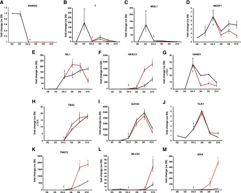
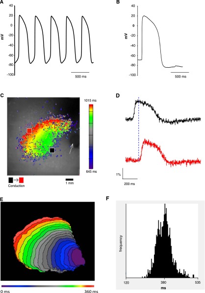
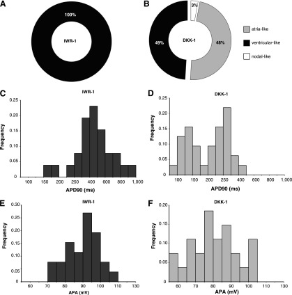
The generation of human ventricular cardiomyocytes from human embryonic stem cells and/or induced pluripotent stem cells could fulfill the demand for therapeutic applications and in vitro pharmacological research; however, the production of a homogeneous population of ventricular cardiomyocytes remains a major limitation. By combining small molecules and growth factors, we developed a fully chemically defined, directed differentiation system to generate ventricular-like cardiomyocytes (VCMs) from human embryonic stem cells and induced pluripotent stem cells with high efficiency and reproducibility. Molecular characterization revealed that the differentiation recapitulated the developmental steps of cardiovascular fate specification. Electrophysiological analyses further illustrated the generation of a highly enriched population of VCMs. These chemically induced VCMs exhibited the expected cardiac electrophysiological and calcium handling properties as well as the appropriate chronotropic responses to cardioactive compounds. In addition, using an integrated computational and experimental systems biology approach, we demonstrated that the modulation of the canonical Wnt pathway by the small molecule IWR-1 plays a key role in cardiomyocyte subtype specification. In summary, we developed a reproducible and efficient experimental platform that facilitates a chemical genetics-based interrogation of signaling pathways during cardiogenesis that bypasses the limitations of genetic approaches and provides a valuable source of ventricular cardiomyocytes for pharmacological screenings as well as cell replacement therapies.

Keywords: Cardiac; Differentiation; Embryonic stem cells; Pluripotent stem cells.


Figures

Figure 1.
Figure 1.
Molecular analysis during the course of the differentiation. Gene expression was analyzed on embryoid bodies at the indicated time points for genes associated with pluripotency (A), mesoderm (B), primitive streak-like (C), cardiac mesoderm (D), cardiac progenitors (E–J), and terminally differentiated cardiomyocytes (K–M). Red line: IWR-1 mediated; black line: dimethyl sulfoxide control. Arrow indicates the initiation of stage 2 of the differentiation. Gene expression was normalized to the housekeeping gene B2M. Values represent means ± SE of a minimum of four independent experiments. #, p < .05 versus D0; ∗, p < .05 IWR-1 versus dimethyl sulfoxide. Abbreviation: D, day.
Figure 2.
Figure 2.
Transcriptional profiling and system biology analyses at the initiation of stage 2. (A): The differentially expressed genes in IWR-1 compared with dimethyl sulfoxide-treated cells were clustered by expression pattern at the indicated time points. (B): The up- and downregulated genes at the indicated time points were analyzed for gene ontology (GO) biological process enrichment. (C): The top 10 transcription factors as putative upstream regulators at each time point were identified with the Expression2Kinases suite. (D): The predicted transcription factors of each time point were used as seed nodes to generate transcription factor networks. The protein-protein interaction network at 4 hours is shown. (E): The transcription factor regulatory networks were analyzed for GO process enrichment. Only signaling processes (i.e., subprocesses of the GO processes “signaling” and “regulation of signaling”) are shown. Abbreviation: dADP, deoxyadenosine diphosphate.
Figure 3.
Figure 3.
Quantification of the cardiomyocyte differentiation efficiency. (A–C): Flow cytometry analysis of the differentiation efficiency of at 21 days after differentiation. Representative contour plots of cells immune labeled with antibodies against the IgG isotype control antibody (A) or the cardiomyocyte marker genes TNNT2 (B) or ACTN2 (C). Values represent means ± SE of six independent differentiation experiments. Alexa Fluor-647 indicates the secondary antibody fluorescence. (D, E): Structural organization of cardiomyocytes. Representative immunofluorescence staining images of the cardiomyocyte-specific marker TNNT2 (red); DNA was counterstained with 4′,6-diamidino-2-phenylindole (blue) (D). The high magnification of the indicated region shows that the cardiomyocytes have well-organized myofilament structures (E). Scale bar = 20 μm. (F, G): Comparison of the cardiomyocyte differentiation efficiency in the current protocol (IWR-1) and the protocol described by Yang et al. [6] (DKK1). Flow cytometric quantification of the percentage of TNNT2-expressing cells treated with IWR-1 or DKK1 or in the absence of either inhibitor (DMSO) (F). Quantitative reverse transcription polymerase chain reaction expression analysis of the cardiomyocyte marker genes NKX2.5, TNNT2, MLC2v, and IRX4 (G). Gene expression was normalized to B2M endogenous control. Values represent means ± SE of a minimum of four independent experiments. ∗, p < .001. Abbreviations: DMSO, dimethyl sulfoxide; SSC, side scatter.
Figure 4.
Figure 4.
Electrophysiological characterization. The action potential (AP) properties of single cells were analyzed using the patch-clamp method. (A, B): Representative AP waveforms of spontaneous (A) and electrically stimulated (B) cells indicating a ventricular-like phenotype. Electrophysiological properties at the multicellular level: Single-cell preparations were plated at high density to form a monolayer and were stained with the voltage-sensitive dye di-4-ANNEPS for high-resolution optical mapping. (C, D): Representative AP tracings (D), which were mapped from two sites distal to the unipolar pacing electrode (indicated by the arrow) that correlates with the two designated points (black and red) in the representative pseudocolor repolarization map recorded from a monolayer (C). The conduction velocity was calculated based on the distance between the two points and the conduction time delay, yielding an overall speed of 2.15 ± 0.35 cm per second (mean ± SE of five independent experiments). (E): Representative isochrones map with 18-ms intervals shows a circular spreading pattern of the optically mapped transmembrane potentials. (F): Histogram shows the distribution of the AP duration at 90% repolarization values that were calculated from 755 spatially distinct locations from five monolayers.
Figure 5.
Figure 5.
Electrophysiological properties of the cardiomyocytes generated with two different protocols. Directed cardiomyocyte differentiation experiments were performed with the current protocol (IWR-1) and the method described by Yang et al. [6] (DKK1). (A, B): Doughnut charts showing the proportion of cardiomyocytes that were classified as atrial-, ventricular- and nodal-like subtypes in the IWR-1 protocol (n = 26) (A) and the DKK1 protocol (n = 31) (B). All cells in the IWR-1 protocol were classified as ventricular-like, whereas the DKK1 protocol generated a heterogeneous population consisting of atrial-like (48%), ventricular-like (49%), and nodal-like cardiomyocytes (3%). (C–F): Frequency distributions of the individual action potential values obtained from single cells in the indicated protocols. Frequency distribution of the APD90 parameters (C, D) and the APA parameters (E, F). The distributions were significantly different in the IWR-1 protocol when compared with the DKK1 protocol (APD90: p = .001; APA: p = .04; Kolmogorov-Smirnov test). Abbreviations: APA, action potential amplitude; APD90, action potential duration at 90% repolarization.
Figure 6.
Figure 6.
Functional characterization of the chemically induced VCMs (ciVCMs). (A, B): Intracellular calcium ([Ca2+]i) transient recordings in ciVCMs. Single-cell preparations were loaded with a fluorescent calcium-sensitive dye (Fluo-4), and calcium transients were recorded in a spinning disk laser confocal microscope utilizing the line-scan mode. Representative [Ca2+]i transient line-scan tracing recorded from an electrically induced (0.2 Hz) ciVCM (A) and during the response to rapid administration of 10 mM of caffeine (B). (C–E): Effect of caffeine application (10 mM) on [Ca2+]i transient parameters. Analyses of [Ca2+]i transient amplitude (C), upstroke velocity (D), and upstroke decay velocity (E). Values represent means ± SE of n = 28 (baseline) and n = 13 (caffeine). ∗, p < .05 versus the baseline values. (F–J): Chronotropic responses of ciVCMs to cardioactive compounds: representative extracellular field potential recordings at baseline (F), after administration of 1 μM of sotalol (G), and 5 μM of isoproterenol (H). Dose-response histograms showing the percentage of change in spontaneously beating rate on administration of escalating concentrations (0.01 μM, 0.1 μM, and 1 μM; n = 12) of sotalol (I) and of isoproterenol (0.05 μM, 0.5 μM, and 5 µM; n = 12) (J), relative to baseline conditions (100%). Values represent means ± SE. #, p < .01 versus the baseline values. Abbreviation: F/F0, fluorescence normalized to baseline fluorescence.
Figure 7.
Figure 7.
Schematic representation of the directed differentiation protocol in two stages. In stage one (days 0–4.5), the hESCs grown in feeder-independent conditions were differentiated toward a multipotent cardiovascular progenitor population by the combinatorial activation of the BMP and nodal/activin signaling pathways. In stage 2 (days 4.5–8), the uncommitted progenitors were terminally differentiated toward ventricular-like cardiomyocytes by the inhibition of the WNT signaling pathway with the small molecule IWR-1. Abbreviations: BMP, bone morphogenic proteins; hESC, human embryonic stem cell.

References

    1. Evans SM, Yelon D, Conlon FL, et al. Myocardial lineage development. Circ Res. 2010;107:1428–1444. - PMC - PubMed
    1. Olson EN. Gene regulatory networks in the evolution and development of the heart. Science. 2006;313:1922–1927. - PMC - PubMed
    1. Noseda M, Peterkin T, Simões FC, et al. Cardiopoietic factors: Extracellular signals for cardiac lineage commitment. Circ Res. 2011;108:129–152. - PubMed
    1. Kehat I, Kenyagin-Karsenti D, Snir M, et al. Human embryonic stem cells can differentiate into myocytes with structural and functional properties of cardiomyocytes. J Clin Invest. 2001;108:407–414. - PMC - PubMed
    1. Laflamme MA, Chen KY, Naumova AV, et al. Cardiomyocytes derived from human embryonic stem cells in pro-survival factors enhance function of infarcted rat hearts. Nat Biotechnol. 2007;25:1015–1024. - PubMed

Publication types

MeSH terms