Review
. 2013:2013:150952.
doi: 10.1155/2013/150952. Epub 2013 Nov 12.

Small-molecule theranostic probes: a promising future in neurodegenerative diseases

Affiliations
Review

Small-molecule theranostic probes: a promising future in neurodegenerative diseases

Suzana Aulić et al. Int J Cell Biol. 2013.

Abstract

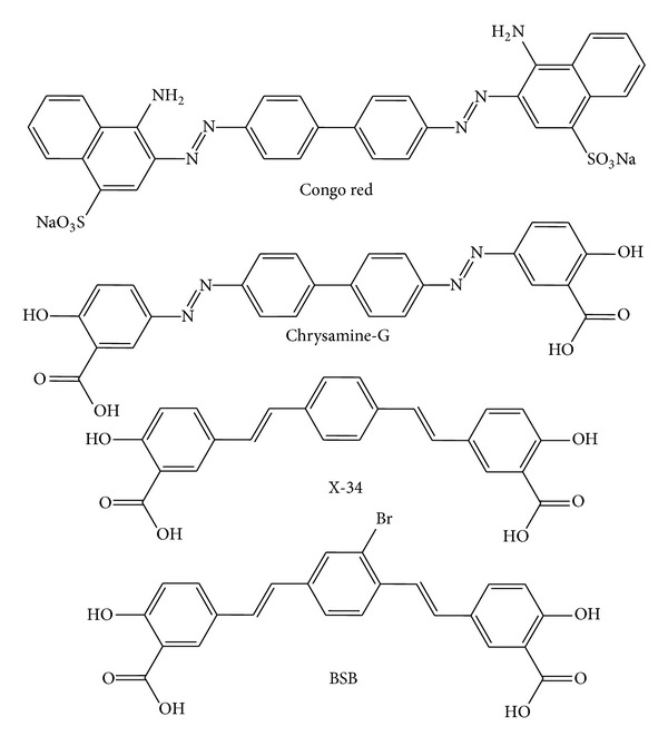
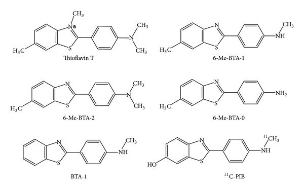
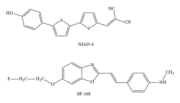
Prion diseases are fatal neurodegenerative illnesses, which include Creutzfeldt-Jakob disease in humans and scrapie, chronic wasting disease, and bovine spongiform encephalopathy in animals. They are caused by unconventional infectious agents consisting primarily of misfolded, aggregated, β -sheet-rich isoforms, denoted prions, of the physiological cellular prion protein (PrP(C)). Many lines of evidence suggest that prions (PrP(Sc)) act both as a template for this conversion and as a neurotoxic agent causing neuronal dysfunction and cell death. As such, PrP(Sc) may be considered as both a neuropathological hallmark of the disease and a therapeutic target. Several diagnostic imaging probes have been developed to monitor cerebral amyloid lesions in patients with neurodegenerative disorders (such as Alzheimer's disease, Parkinson's disease, and prion disease). Examples of these probes are Congo red, thioflavin T, and their derivatives. We synthesized a series of styryl derivatives, denoted theranostics, and studied their therapeutic and/or diagnostic potentials. Here we review the salient traits of these small molecules that are able to detect and modulate aggregated forms of several proteins involved in protein misfolding diseases. We then highlight the importance of further studies for their practical implications in therapy and diagnostics.


Figures

Figure 1
Figure 1
Pathogenic mutations and polymorphisms in the human PrP. The pathogenic mutations associated with human prion diseases are shown above the human PrP coding sequence. These consist of 1, 2, or 4–9 octapeptide repeat insertions (OPR1-9) within the octapeptide repeat region between codons 51 and 91, a 2 octapeptide repeat deletion (OPR2), and various point mutations causing missense or stop amino-acid substitutions. Point mutations are designated by the wild-type amino acid preceding the codon number, followed by the mutant residue, using single letter amino-acid nomenclature. Polymorphic variants are shown below the PrP coding sequence.
Figure 2
Figure 2
(a) The “template-assistance model” [8] and (b) the “seeding nucleation model” [9].
Figure 3
Figure 3
(a) A 50-year-old man with definite sCJD. Axial DWI shows pathologic hyperintensity in bilateral posterior temporoparietal neocortex. Cortex along parietal-occipital fissure is abnormally hyperintense (vertical arrows), but primary visual region is spared (horizontal arrows). Note asymmetric abnormal hyperintensity in right cingulum (arrowhead). Striatum is uninvolved. (b) FLAIR image at same level shows more subtle pathologic hyperintensity in all abnormal regions on DWI, as shown in cingulate cortex (arrowhead) [10]. (c) Definite sCJD (MM1); total duration: 10 months; EEG at 6 weeks: typical (used to classify case as probable); source: http://www.eurocjd.ed.ac.uk.
Figure 4
Figure 4
Detection of PK-sensitive PrPSc. (a) Conventional Western blot of PrP treated with or without PK. No PrP was observed after PK treatment in the samples from non-CJD. The PK-resistant PrP27–30 was indicated in the sample from sCJD. Samples were digested with 50 μg/mL proteinase K for 1 hour at 37°C, completely hydrolyzing PrPC. Proteinase digestion cleaves ~90 amino acids from the amino terminus of PrPSc to generate PrP27–30. Blot is developed with anti-PrP mouse monoclonal antibody 3F4 [11].
Figure 5
Figure 5
PET imaging of human AD brain using [11C-] PIB [12]. PIB standardized uptake value (SUV) images show a marked difference between PIB retention in AD patients and healthy control subjects. PET images of a 67-year-old healthy control subject (left) and a 79-year-old AD patient. The left column shows lack of PIB retention in the entire gray matter of the healthy subject.
Figure 6
Figure 6
(a) Chemical formula of the three promising antiprionic compounds and in vitro staining with (b) STB-8 compound (β-amyloid plaques) and (c) G8 compound (PrPSc deposits).
Scheme 1
Scheme 1
Chemical structures of Congo red and its derivatives, potential in vivo imaging agents for β-amyloid plaques.
Scheme 2
Scheme 2
Chemical structures of thioflavin T and its derivatives, useful for potential in vivo imaging for β-amyloid plaques.
Scheme 3
Scheme 3
Chemical structures of potential in vivo imaging agents for β-amyloid plaques.

Similar articles

Cited by

References

    1. Rowe CC, Ackerman U, Browne W, et al. Imaging of amyloid β in Alzheimer’s disease with 18F-BAY94-9172, a novel PET tracer: proof of mechanism. The Lancet Neurology. 2008;7(2):129–135. - PubMed
    1. O'keefe GJ, Saunder TH, Ng S, et al. Radiation dosimetry of beta-amyloid tracers 11c-Pib and 18f-bay94-9172. Journal of Nuclear Medicine. 50:309–315. - PubMed
    1. Koole M, Lewis DM, Buckley C, et al. Whole-body biodistribution and radiation dosimetry of 18F-GE067: a radioligand for in vivo brain amyloid imaging. Journal of Nuclear Medicine. 2009;50(5):818–822. - PubMed
    1. Fernandez-Fernandez A, Manchanda R, McGoron AJ. Theranostic applications of nanomaterials in cancer: drug delivery, image-guided therapy, and multifunctional platforms. Applied Biochemistry and Biotechnology. 2011;165(7-8):1628–1651. - PMC - PubMed
    1. Lobatto ME, Fuster V, Fayad ZA, Mulder WJM. Perspectives and opportunities for nanomedicine in the management of atherosclerosis. Nature Reviews Drug Discovery. 2011;10(11):835–852. - PMC - PubMed