

Sinus fungus ball in the Japanese population: clinical and imaging characteristics of 104 cases
- PMID: 24324499
- PMCID: PMC3845720
- DOI: 10.1155/2013/731640
Sinus fungus ball in the Japanese population: clinical and imaging characteristics of 104 cases
Abstract
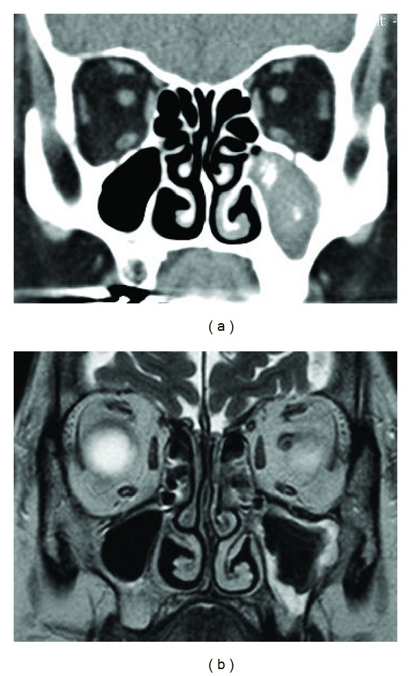
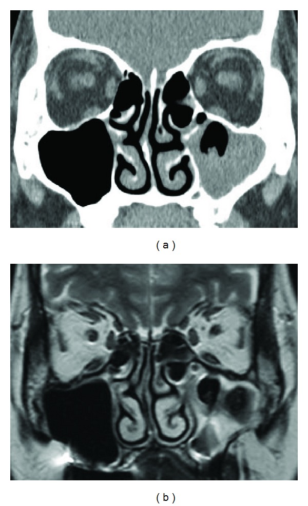
Sinus fungus ball is defined as noninvasive chronic fungal rhinosinusitis occurring in immunocompetent patients with regional characteristics. The clinical and imaging characteristics of paranasal sinus fungus ball were retrospectively investigated in 104 Japanese patients. All patients underwent endoscopic sinus surgery. Preoperative computed tomography (CT), magnetic resonance (MR) imaging, age, sex, chief complaint, causative fungus, and clinical outcome were analyzed. Patients were aged from 25 to 79 years (mean 58.8 years). Female predominance was noted (58.7%). Most common symptoms were nasal discharge and facial pain. CT showed high density area in 82.0% of the cases (82/100), whereas T2-weighted MR imaging showed low intensity area in 100% of the cases (32/32). Histological examination showed that most causative agents were Aspergillus species (94.2% (98/104)). Culture test was positive for 16.7% (11/66). Recurrence was found in 3.2% (3/94). Older age and female predominance were consistent with previous reports. MR imaging is recommended to confirm the diagnosis.
Figures
References
-
- Grosjean P, Weber R. Fungus balls of the paranasal sinuses: a review. European Archives of Oto-Rhino-Laryngology. 2007;264(5):461–470. - PubMed
-
- Nicolai P, Lombardi D, Tomenzoli D, et al. Fungus ball of the paranasal sinuses: experience in 160 patients treated with endoscopic surgery. Laryngoscope. 2009;119(11):2275–2279. - PubMed
-
- Dufour X, Kauffmann-Lacroix C, Ferrie JC, Goujon JM, Rodier MH, Klossek JM. Paranasal sinus fungus ball: epidemiology, clinical features and diagnosis. A retrospective analysis of 173 cases from a single medical center in France, 1989–2002. Medical Mycology. 2006;44(1):61–67. - PubMed
-
- Ferreiro JA, Carlson BA, Cody DT., III Paranasal sinus fungus balls. Head and Neck. 1997;19(6):481–486. - PubMed
-
- Klossek J-M, Serrano E, Péloquin L, Percodani J, Fontanel J-P, Pessey J-J. Functional endoscopic sinus surgery and 109 mycetomas of paranasal sinuses. Laryngoscope. 1997;107(1):112–117. - PubMed
LinkOut - more resources
Full Text Sources
Other Literature Sources
