. 2013 Dec 4;8(12):e80768.
doi: 10.1371/journal.pone.0080768. eCollection 2013.

Glycation accelerates fibrillization of the amyloidogenic W7FW14F apomyoglobin

Affiliations

Glycation accelerates fibrillization of the amyloidogenic W7FW14F apomyoglobin

Clara Iannuzzi et al. PLoS One. .

Retraction in

Abstract

Neurodegenerative diseases are associated with misfolding and deposition of specific proteins, either intra or extracellularly in the nervous system. Advanced glycation end products (AGEs) originate from different molecular species that become glycated after exposure to sugars. Several proteins implicated in neurodegenerative diseases have been found to be glycated in vivo and the extent of glycation is related to the pathologies of the patients. Although it is now accepted that there is a direct correlation between AGEs formation and the development of neurodegenerative diseases, several questions still remain unanswered: whether glycation is the triggering event or just an additional factor acting on the aggregation pathway. To this concern, in the present study we have investigated the effect of glycation on the aggregation pathway of the amyloidogenic W7FW14F apomyoglobin. Although this protein has not been related to any amyloid disease, it represents a good model to resemble proteins that intrinsically evolve toward the formation of amyloid aggregates in physiological conditions. We show that D-ribose, but not D-glucose, rapidly induces the W7FW14F apomyoglobin to generate AGEs in a time-dependent manner and protein ribosylation is likely to involve lysine residues on the polypeptide chain. Ribosylation of the W7FW14F apomyoglobin strongly affects its aggregation kinetics producing amyloid fibrils within few days. Cytotoxicity of the glycated aggregates has also been tested using a cell viability assay. We propose that ribosylation in the W7FW14F apomyoglobin induces the formation of a cross-link that strongly reduces the flexibility of the H helix and/or induce a conformational change that favor fibril formation. These results open new perspectives for AGEs biological role as they can be considered not only a triggering factor in amyloidosis but also a player in later stages of the aggregation process.


Conflict of interest statement

Competing Interests: The authors have declared that no competing interests exist.

Figures

Figure 1
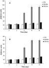
Figure 1. Glycation of the W7FW17F apomyoglobin.
Protein glycation monitored by fluorescence spectroscopy. W7FW14F apomyoglobin was incubated in the absence (white bar) and in the presence of 0.5 M D-ribose (grey bar) and 0.5 M D-glucose (black bar) and changes in maximal fluorescence intensity were monitored at different time intervals. Protein concentration was 40 µM, other experimental details are described in the Materials and Methods section. (A) Maximal fluorescence intensity recorded at 410 nm upon excitation at 320 nm. (B) Maximal fluorescence intensity recorded at 425 nm upon excitation at 370 nm. The average value (±SD) of a quadruplicate experiment is plotted.
Figure 2
Figure 2. Western blot of ribosylated W7FW17F apomyoglobin.
W7FW14F apomyoglobin was incubated in the absence and in the presence of 0.5M D-ribose and aliquots were taken at different time intervals. (A) Western blot analysis using an anti-myoglobin antibody. (B) Dot blot analysis using an anti AGE antibody. Experimental conditions are described in the Materials and Methods section.
Figure 3
Figure 3. Effect of ribosylation on the amyloid formation for the W7FW17F apomyoglobin.
W7FW14F apomyoglobin (40 µM) was incubated in the absence (light gray bar) and in the presence of 0.5M D-ribose (dark grey bar) and aliquots of each sample at different incubation time intervals were monitored by ThT fluorescence. The average value (±SD) of a quadruplicate experiment is plotted.
Figure 4
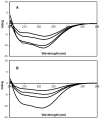
Figure 4. Effect of ribosylation on the CD activity of the W7FW17F apomyoglobin.
Time dependence of the far-UV CD activity of the amyloid forming W7FW14F apomyoglobin mutant at pH 7.0 in the absence (A) and in the presence of 0.5 M D-ribose (B). From the lower to the upper spectrum, times are: 1, 60, 180, and 360 min. Protein concentration was 20 µM.
Figure 5
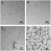
Figure 5. Effect of ribosylation on the morphology of the W7FW17F apomyoglobin aggregates.
Electron microscopy images of the W7FW14F apomyoglobin in the absence of D-ribose at the beginning (A) and after 5 days of incubation (B), and in the presence of D-ribose at the beginning (C) and after 5 days of incubation (D). Experimental conditions are described in the Materials and Methods section.
Figure 6
Figure 6. Solvent accessible regions in the wild-type and W7FW17F apomyoglobin.
Schematic representation of the A-H helices in the wild-type and W7FW14F apomyoglobin. Solvent accessible helices for both protein are shown in grey. Experimental conditions are described in .
Figure 7
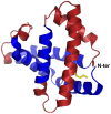
Figure 7. Proposed cross-linking in the glycated W7FW17F apomyoglobin.
Structural representation of the W7FW14F apomyoglobin modeled on the wild-type protein X-ray structure (pdb 2JHO). Regions involved in the fibril core are shown in dark blue while solvent accessible regions are shown in red. Glycation is likely to promote a cross-linking between the N-terminus and the side chain of Lys133 (shown in yellow). The results are visualized using PyMol (The PyMOL Molecular Graphics System, Version 1.5.0.4 Schrodinger, LLC).
Figure 8
Figure 8. Effect of ribosylation on the cytotoxicity of the W7FW17F apomyoglobin aggregates.
NIH-3T3 cells were exposed to aggregates formed in the absence (light grey) and in the presence of D-ribose (dark grey). Aliquots of protein were taken at 0, 5 and 15 days from the onset of aggregation and incubated for 24 h with cells. Data are expressed as average percentage of MTT reduction ±SD relative to cell treated with medium alone or medium plus D-ribose, from triplicate wells from 5 separate experiments (p<0.01). The white bar represents the negative control (cells treated with medium plus 0.25 M D-ribose). Other experimental conditions are described in the Materials and Methods section.

References

    1. Dobson CM (2003) Protein folding and misfolding. Nature 426: 884–890. - PubMed
    1. Luheshi LM, Dobson CM (2009) Bridging the gap: from protein misfolding to protein mis-folding diseases. FEBS Letters 583: 2581–2586. - PubMed
    1. Serpell LC, Sunde M, Benson MD, Tennent GA, Pepys MB, et al. (2000) The protofilament substructure of amyloid fibrils. J Mol Biol 300: 1033–1039. - PubMed
    1. Kodali R, Wetzel R (2007) Polymorphism in the intermediates and products of amyloid assembly. Current Opinion in Structural Biology 17: 48–57. - PubMed
    1. Invernizzi G, Papaleo E, Sabate R, Ventura S (2012) Protein aggregation: mechanisms and functional consequences. Int J Biochem Cell Biol. 44: 1541–1554. - PubMed

Publication types

MeSH terms

LinkOut - more resources