. 2013 Dec 4;8(12):e81158.
doi: 10.1371/journal.pone.0081158. eCollection 2013.

Canonical Wnt/β-catenin signalling is essential for optic cup formation

Affiliations

Canonical Wnt/β-catenin signalling is essential for optic cup formation

Anna-Carin Hägglund et al. PLoS One. .

Abstract

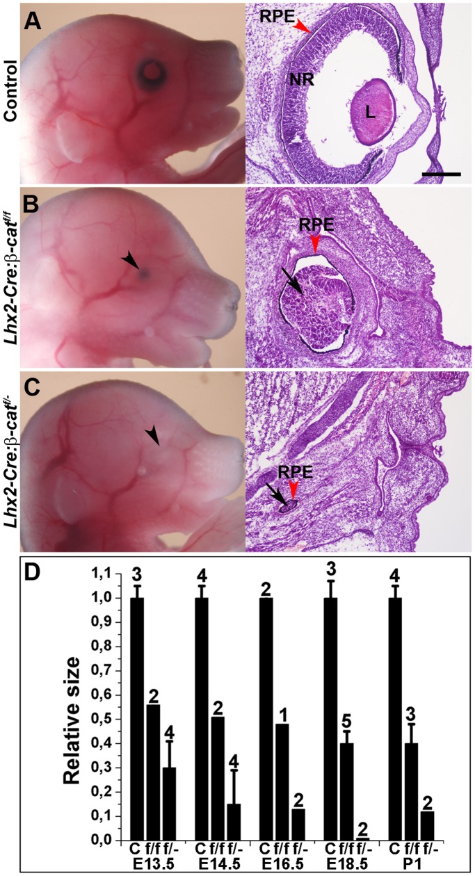
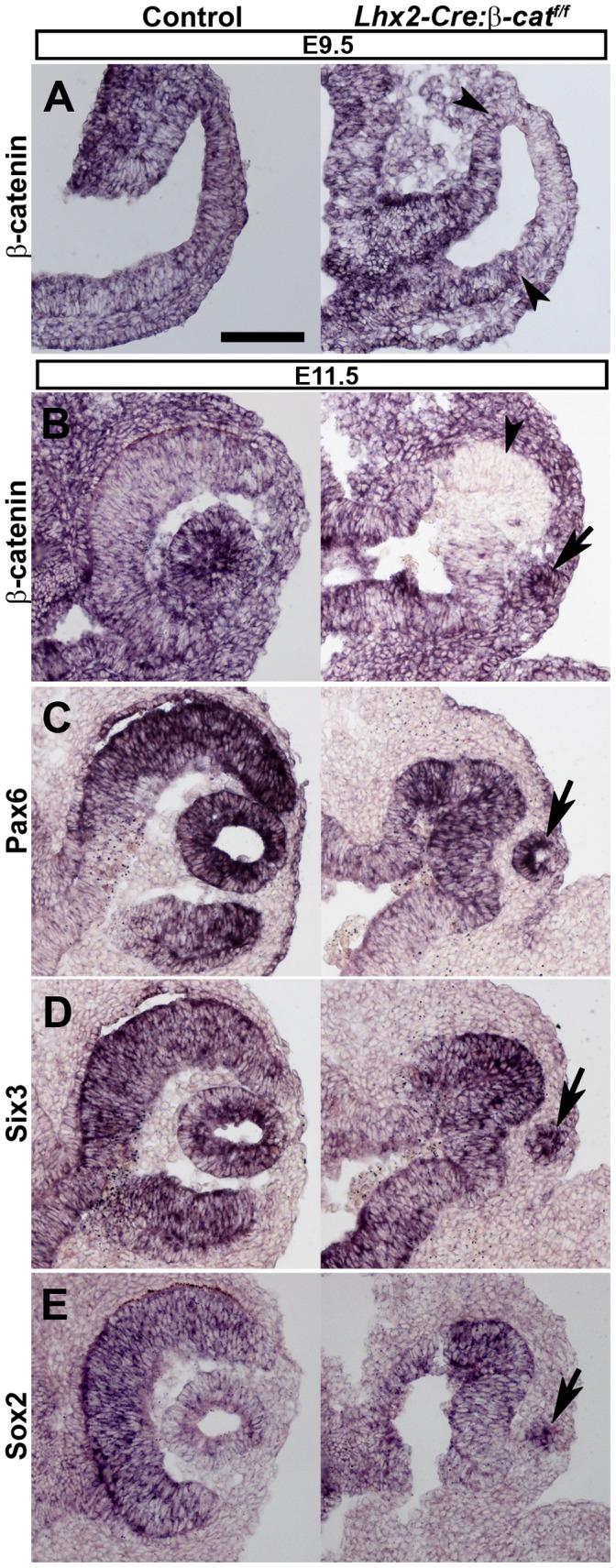
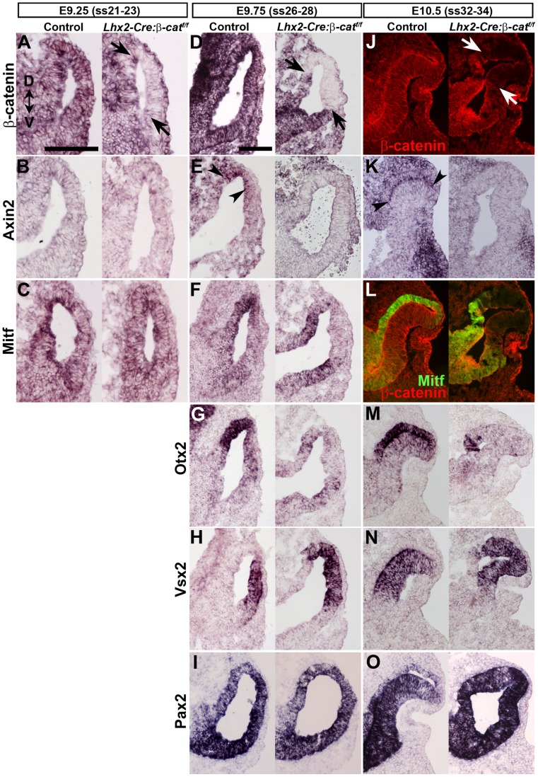
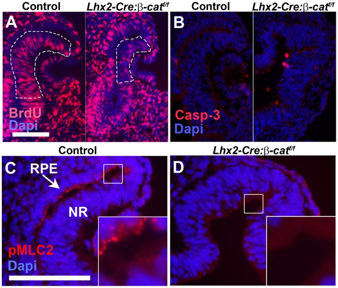
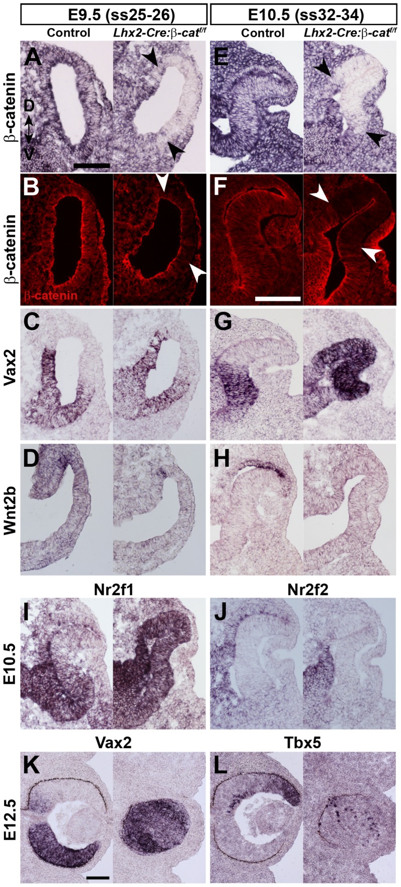
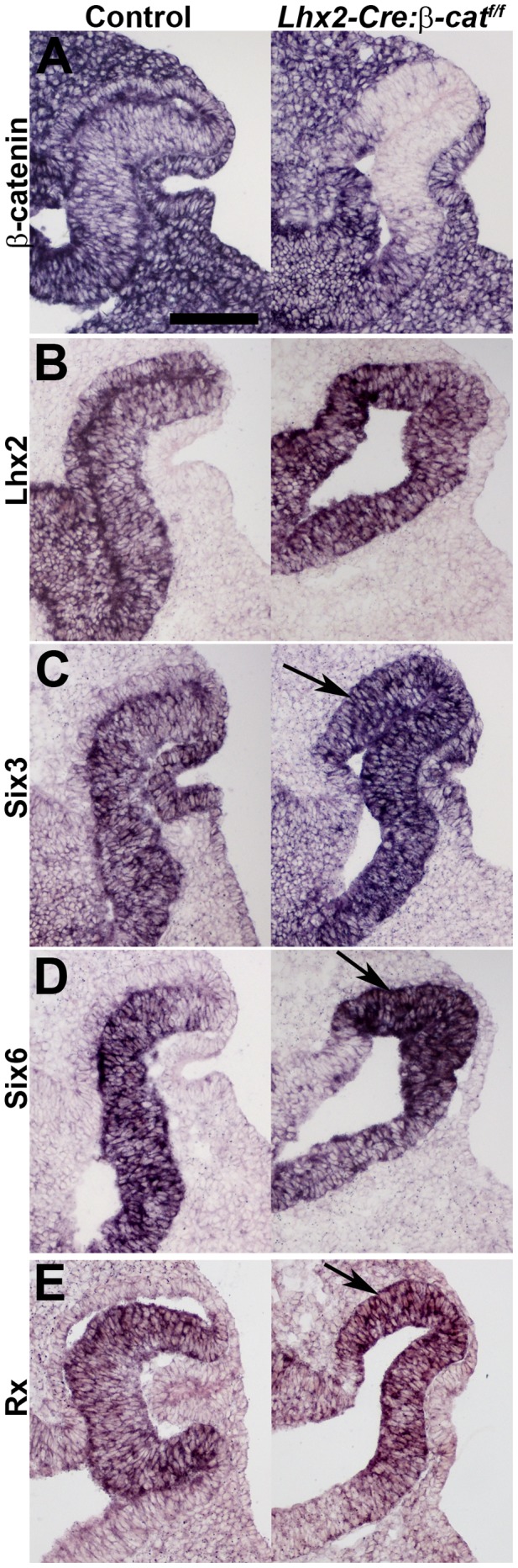
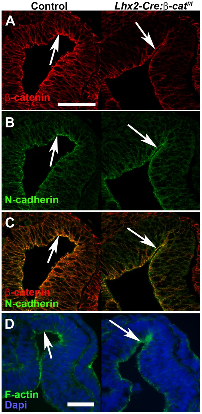
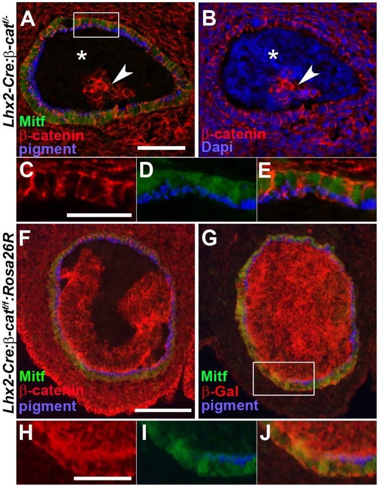
A multitude of signalling pathways are involved in the process of forming an eye. Here we demonstrate that β-catenin is essential for eye development as inactivation of β-catenin prior to cellular specification in the optic vesicle caused anophthalmia in mice. By achieving this early and tissue-specific β-catenin inactivation we find that retinal pigment epithelium (RPE) commitment was blocked and eye development was arrested prior to optic cup formation due to a loss of canonical Wnt signalling in the dorsal optic vesicle. Thus, these results show that Wnt/β-catenin signalling is required earlier and play a more central role in eye development than previous studies have indicated. In our genetic model system a few RPE cells could escape β-catenin inactivation leading to the formation of a small optic rudiment. The optic rudiment contained several neural retinal cell classes surrounded by an RPE. Unlike the RPE cells, the neural retinal cells could be β-catenin-negative revealing that differentiation of the neural retinal cell classes is β-catenin-independent. Moreover, although dorsoventral patterning is initiated in the mutant optic vesicle, the neural retinal cells in the optic rudiment displayed almost exclusively ventral identity. Thus, β-catenin is required for optic cup formation, commitment to RPE cells and maintenance of dorsal identity of the retina.


Conflict of interest statement

Competing Interests: The authors have declared that no competing interests exist.

Figures

Figure 1
Figure 1. β-catenin mutant embryos are anophthalmic.
(A–C) The left panels show lateral views of E16.5 embryos of control (A), Lhx2-Cre:β-cateninflox/flox (β-catf/f) (B) and Lhx2-Cre:β-cateninflox/ (β-catf/ ) (C) embryos. Arrow heads indicate where the eye should be located in the mutant embryos. The right panels show hematoxylin/eosin staining of coronal tissue sections of the same embryos. Black arrows indicate the optic rudiment surrounded by RPE cells (red arrow heads) that can develop in the mutant embryos. NR: neural retina. L: lens. RPE: retinal pigment epithelium. (D) Relative size of the optic vesicle-derived structure in the Lhx2-Cre:β-cateninflox/flox (f/f) embryos and Lhx2-Cre:β-cateninflox/ (f/−) embryos compared with control embryos (c) at the indicated embryonic age and postnatal day 1 (P1). To get an approximate value of the relative eye size for the different genotypes, the largest diameter of the control eyes and the mutant optic rudiment at different ages were measured and compared. The diameter of the eye in control embryos at each developmental stage is arbitrarily defined as 1.0. The number of eyes analysed for each genotype at the respective age is indicated for each group. SD is indicated when applicable. Scale bar: 200 µm.
Figure 2
Figure 2. β-catenin mutant optic vesicles do not form an optic cup.
(A) In situ hybridization analyses of β-catenin expression on coronal sections of optic vesicles of an E9.5 control embryo (left panel) and an Lhx2-Cre:β-cateninflox/flox embryo (right panel). The arrow heads indicate the boundaries of the area in the optic vesicle where β-catenin has been inactivated. (B–E) In situ hybridization analyses for gene expression of the indicated genes on coronal sections of control embryos (left panels) and Lhx2-Cre:β-cateninflox/flox embryos (right panels) at E11.5. Arrows indicate the lens pit where the early lens ectoderm remains attached to the epidermal ectoderm in mutants. Arrow head in (B) indicate the area where the β-catenin gene has been inactivated. Scale bar: 100 µm.
Figure 3
Figure 3. Canonical Wnt/β-catenin signalling is required for RPE cell commitment.
(A–C) In situ hybridization analyses of the indicated genes on coronal sections of E9.25 somite stage 21–23 (ss21–23) control embryos (left panels) and Lhx2-Cre:β-cateninflox/flox embryos (right panels). (D–I) In situ hybridization analyses of the indicated genes on coronal sections of E9.75 (ss26–28) control embryos (left panels) and Lhx2-Cre:β-cateninflox/flox embryos (right panels). (J–O) In situ hybridization analyses (K, M–O) or immunohistochemical analyses (J, L) for gene expression of the indicated genes on coronal sections of E10.5 (ss32–34) control embryos (left panels) and Lhx2-Cre:β-cateninflox/flox embryos (right panels). β-catenin protein is indicated by red labelling in J and L while Mitf protein is indicated by green labelling in L. Arrows indicate the area where β-catenin has been inactivated in A, D and J. Arrow heads indicate the area of Axin2 expression in E and K. Dorsal-ventral (D–V) orientation for all panels is indicated in A. Scale bars: (A–C and D–O) 100 µm.
Figure 4
Figure 4. Decreased proliferation and change in physical properties of mutant optic vesicle.
(A) Immunohistochemical analysis for the presence of BrdU-labelled cells in the prospective neural retina on a coronal section of an E10.5 control embryo (left panel) and an Lhx2-Cre:β-cateninflox/flox embryo (right panel). The part of the prospective neural retina that was analysed for BrdU+ cells in the control and mutant embryos are outlined. (B) Immunohistochemical analysis for the presence apoptotic cells as revealed by the presence of activated Casp-3+ cells on coronal section of an E10.5 control embryo (left panel) and a Lhx2-Cre:β-cateninflox/flox embryo (right panel). (C) Immunohistochemical analyses for the presence of phosphorylated myosin light chain 2 (pMLC2) on a coronal section of an E10.5 control embryo. (D) Immunohistochemical analyses for the presence of pMLC2 on a coronal section on an E10.5 Lhx2-Cre:β-cateninflox/flox embryo. Insets in C and D are a magnification of the areas indicated by the squares. Scale bars: (A, B and C, D) 100 µm.
Figure 5
Figure 5. β-catenin is important for maintenance of dorsoventral patterning of the retina.
(A–D) In situ hybridization analyses (A, C, D) and immunohistochemical analysis (B) on coronal sections of E9.5 (ss25–26) optic vesicles from control (left panels) and on Lhx2-Cre:β-cateninflox/flox embryos (right panels). (E–H) In situ hybridization analyses (E, G, H) and immunohistochemical analysis (F) on coronal sections of E10.5 (ss32–34) optic cups from control (left panels) and Lhx2-Cre:β-cateninflox/flox embryos (right panels). Black arrow heads in A and E, and white arrow heads in B and F, indicate the boundaries of the area where β-catenin has been inactivated. (I, J) In situ hybridization analyses on coronal sections of E10.5 (ss32–34) optic cups from control (left panel) and Lhx2-Cre:β-cateninflox/flox embryos (right panels). (K, L) In situ hybridization analyses on coronal sections of an eye from E12.5 control (left panels) and Lhx2-Cre:β-cateninflox/flox embryos. Dorsal-ventral (D–V) orientation of all panels is indicated in A. Scale bars: (A–E, G–J, F and K, L) 100 µm.
Figure 6
Figure 6. Dorsal shift in expression of retina-specific eye field transcription factors in the mutant optic vesicle.
(A–E) In situ hybridization analyses on coronal sections of E10.5 optic cups from control embryos (left panels) and Lhx2-Cre:β-cateninflox/flox mutant embryos (right panels). Arrows indicate the dorsal shift in expression of the retina-specific transcription factors Six3, Six6 and Rx in mutant embryos (C–E). Scale bar: 100 µm.
Figure 7
Figure 7. β-catenin remains associated with N-cadherin and F-actin at the apical side of the cells in the mutant optic vesicle.
(A–D) Immunohistochemical analyses for cellular localisation of the indicated proteins on coronal sections of E10 (ss30–31) optic vesicles from control embryos (left panels) and Lhx2-Cre:β-cateninflox/flox embryos (right panels). (C) is a merge of (A) and (B) to show co-localisation (yellow) in the apical side of the cells in the optic vesicle on both control and mutant embryos (arrows). (D) A serial section of the same embryo as in (A–C) revealing F-actin protein at the apical side of the cells in both control and mutant optic vesicles (arrows). Scale bars: (A–C and D) 50 µm.
Figure 8
Figure 8. The few RPE cells that develop in the mutant embryos are derived from eye committed progenitor cells in the anterior neural plate that escape β-catenin inactivation.
(A) Immunohistochemical analyses of Mitf (green) and β-catenin (red) on coronal sections of an E13.5 optic rudiment in an Lhx2-Cre:β-cateninflox/ embryo. RPE cells are identified by pigment (pseudocoloured blue) and Mitf (green). (B) Immunohistochemical analysis of β-catenin and DAPI labelling of the same coronal section as in (A) to reveal that both β-catenin+ and β-catenin cells are present in the optic rudiment that develops in mutant embryos. * indicates the area of β-catenin non-RPE cells and the arrow head indicates the area of β-catenin+ non-RPE cells in the optic rudiment. (C–E) is a magnification of the area indicated by a rectangle in (A). (C) and (D) are merged in (E) to show co-localisation of β-catenin, Mitf and pigment. (F) Immunohistochemical analysis of Mitf (green) and β-catenin (red) on a coronal section of an E12.5 optic rudiment in a Lhx2-Cre:β-cateninflox/flox:ROSA26R embryo. (G) Immunohistochemical analysis of Mitf (green) and β-Gal (red) on a serial section following (F) revealing that all cells in the optic rudiment, including the RPE cells, are β-Gal+ and hence derived from Cre+ progenitor cells in the anterior neural plate. (H–J) is a magnification of the area indicated by a rectangle in (G). (H) and (I) are merged in (J) to show co-localisation of Mitf, β-Gal and pigment. Scale bars: (A, B, and H–J) 50 µm. (C–E) 25 µm. (F, G) 100 µm.

References

    1. Wilson SW, Houart C (2004) Early steps in the development of the forebrain. Dev Cell 6: 167–181. - PMC - PubMed
    1. Kaufman M (1994) The atlas of mouse development. San Diego: Academic Press Limited.
    1. Chow RL, Lang RA (2001) Early eye development in vertebrates. Annu Rev Cell Dev Biol 17: 255–296. - PubMed
    1. Eiraku M, Takata N, Ishibashi H, Kawada M, Sakakura E, et al. (2011) Self-organizing optic-cup morphogenesis in three-dimensional culture. Nature 472: 51–56. - PubMed
    1. Horsford DJ, Nguyen MT, Sellar GC, Kothary R, Arnheiter H, et al. (2005) Chx10 repression of Mitf is required for the maintenance of mammalian neuroretinal identity. Development 132: 177–187. - PubMed

Publication types

MeSH terms

LinkOut - more resources