

New host range for Hematodinium in southern Australia and novel tools for sensitive detection of parasitic dinoflagellates
- PMID: 24324829
- PMCID: PMC3855790
- DOI: 10.1371/journal.pone.0082774
New host range for Hematodinium in southern Australia and novel tools for sensitive detection of parasitic dinoflagellates
Abstract
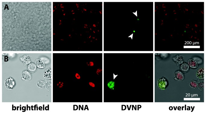
Hematodinium is a parasitic dinoflagellate and emerging pathogen of crustaceans. It preferably manifests in haemolymph of marine decapod crustaceans, killing a large variety of genera with significant impacts on fisheries worldwide. There is, however, evidence that some crustacean stocks harbor high prevalence, low intensity infections that may not result in widespread host mortality and are therefore hard to detect. The most widely used methods for detection of Hematodinium are conventional blood smears and polymerase chain reaction (PCR) against ribosomal RNAs. Blood smears demand a trained investigator, are labor intensive and not readily scalable for high-throughput sampling. PCRs only detect parasite DNA and can also suffer from false negatives and positives. In order to develop alternative detection tools for Hematodinium cells in decapod crustaceans we employed an immunological approach against a newly identified, abundant dinoflagellate-specific nuclear protein--Dinoflagellate/Viral NucleoProtein (DVNP). Both immunofluorescence assay (IFA) and Western blot methods against DVNP showed high sensitivity of detection. The Western blot detects Hematodinium parasites to levels of 25 parasites per milliliter of crustacean haemolymph, with the potential for sample pooling and screening of large samples. Using both PCR and these new tools, we have identified Hematodinium cells present in three new host crab taxa, at high prevalence but with no sign of pathogenesis. This extends the known range of Hematodinium to southern Australia.
Conflict of interest statement
Figures


References
-
- Small HJ, Shields JD, Reece KS, Bateman K, Stentiford GD (2012) Morphological and molecular characterization of Hematodinium perezi (Dinophyceae: Syndiniales), a dinoflagellate parasite of the harbour crab, Liocarcinus depurator . J Eukaryot Microbiol 59: 54–66. doi:10.1111/j.1550-7408.2011.00592.x. PubMed: 22092696. - DOI - PubMed
-
- Hudson D, Shields J (1994) Hematodinium australis n. sp., a parasitic dinoflagellate of the sand crab Portunus pelagicus from Moreton Bay, Australia. Dis Aquat Org 19: 109–119. doi:10.3354/dao019109. - DOI
-
- Field RH, Chapman CJ, Taylor AC, Douglas DMN, Vickerman K (1992) Infection of the Norway lobster Nephrops norvegicus by a Hematodinium-like species of dinoflagellate on the west coast of Scotland. Dis Aquat Org 13: 1–15. doi:10.3354/dao013001. - DOI
-
- Gornik SG, Albalat A, Atkinson RJA, Coombs GH, Neil DM (2010) The influence of defined ante-mortem stressors on the early post-mortem biochemical processes in the abdominal muscle of the Norway lobster, Nephrops norvegicus (Linnaeus, 1758). Mar Biol Res 6: 223–238. doi:10.1080/17451000903147468. - DOI
Publication types
MeSH terms
LinkOut - more resources
Full Text Sources
Other Literature Sources
Molecular Biology Databases
