. 2014 Apr;50(4):825-37.
doi: 10.1165/rcmb.2013-0386OC.

Tumor necrosis factor-α accelerates the resolution of established pulmonary fibrosis in mice by targeting profibrotic lung macrophages

Affiliations

Tumor necrosis factor-α accelerates the resolution of established pulmonary fibrosis in mice by targeting profibrotic lung macrophages

Elizabeth F Redente et al. Am J Respir Cell Mol Biol. 2014 Apr.

Abstract

Idiopathic pulmonary fibrosis (IPF) is a relentless, fibrotic parenchymal lung disease in which alternatively programmed macrophages produce profibrotic molecules that promote myofibroblast survival and collagen synthesis. Effective therapies to treat patients with IPF are lacking, and conventional therapy may be harmful. We tested the hypothesis that therapeutic lung delivery of the proinflammatory cytokine tumor necrosis factor (TNF)-α into wild-type fibrotic mice would reduce the profibrotic milieu and accelerate the resolution of established pulmonary fibrosis. Fibrosis was assessed in bleomycin-instilled wild-type and TNF-α(-/-) mice by measuring hydroxyproline levels, static compliance, and Masson's trichrome staining. Macrophage infiltration and programming status was assessed by flow cytometry of enzymatically digested lung and in situ immunostaining. Pulmonary delivery of TNF-α to wild-type mice with established pulmonary fibrosis was found to reduce their fibrotic burden, to improve lung function and architecture, and to reduce the number and programming status of profibrotic alternatively programmed macrophages. In contrast, fibrosis and alternative macrophage programming were prolonged in bleomycin-instilled TNF-α(-/-) mice. To address the role of the reduced numbers of alternatively programmed macrophages in the TNF-α-induced resolution of established pulmonary fibrosis, we conditionally depleted macrophages in MAFIA (MAcrophage Fas-Induced Apoptosis) mice. Conditional macrophage depletion phenocopied the resolution of established pulmonary fibrosis observed after therapeutic TNF-α delivery. Taken together, our results show for the first time that TNF-α is involved in the resolution of established pulmonary fibrosis via a mechanism involving reduced numbers and programming status of profibrotic macrophages. We speculate that pulmonary delivery of TNF-α or augmenting its signaling pathway represent a novel therapeutic strategy to resolve established pulmonary fibrosis.


Figures

Figure 1.
Figure 1.
The time course of fibrosis development and resolution after bleomycin. (A) Total protein concentration in bronchoalveolar lavage fluid (BALF). (B) Quantification of lavaged bronchoalveolar lavage (BAL) cells after bleomycin-instillation. (C) Hydroxyproline levels measured in saline and bleomycin-instilled mice. (D) Static compliance in saline and bleomycin-instilled mice. (E) Mouse lung sections stained with Masson’s trichrome. Under normal conditions, collagen deposition (blue) is limited to areas adjacent to airways and large vessels but spreads throughout the lung in response to bleomycin instillation. Original magnification: ×20. *P < 0.05, **P < 0.01, and ***P < 0.001.
Figure 2.
Figure 2.
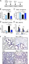
Intratracheal delivery of tumor necrosis factor (TNF)-α after bleomycin-instillation accelerates the resolution of pulmonary fibrosis. (A) Schematic showing the TNF-α treatment regimen given after bleomycin instillation. All mice were harvested at Week 5. (B) TNF-α significantly attenuates the bleomycin-mediated increase in lung hydroxyproline levels (***P < 0.001). (C) Static compliance is significantly reduced after bleomycin instillation and returns to saline levels after TNF-α administration (**P < 0.001). (D and E) Total BALF protein levels (D) and lavaged cells in the BAL (E) are significantly decreased after TNF-α administration (***P < 0.001). (F) Mouse lung sections after TNF-α administration have less collagen staining by Masson’s trichrome compared with bleomycin-instilled mice. Bleo = bleomycin; Sal = saline. Original magnification: ×20.
Figure 3.
Figure 3.
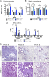
TNF-α−/− mice exhibit delayed resolution of bleomycin-induced pulmonary fibrosis. (A) Hydroxyproline levels remained significantly elevated at 6 and 8 weeks after bleomycin in TNF-α−/− mice compared with wild-type mice (*P < 0.05). (B) Static compliance was significantly decreased at 6 and 8 weeks after bleomycin in TNF-α−/− mice compared with wild-type mice (*P < 0.05). (C) Lavaged cells were significantly elevated in TNF-α−/− mice at 6 and 8 weeks after bleomycin instillation compared with wild-type mice (***P < 0.001) and saline controls (P < 0.001). (D) Masson’s trichrome–stained lung sections from bleomycin-instilled TNF-α−/− mice exhibit impaired resolution of fibrosis and increased inflammation (pink) compared with bleomycin-instilled wild-type mice. Original magnification: ×20.
Figure 4.
Figure 4.
(See figure legend on following page) CD11c+CD11bvarF4/80+ macrophages are alternatively programmed during bleomycin-induced fibrosis. (A) Representative gating strategy to identify pulmonary macrophage subsets from whole lung digest in saline and bleomycin-instilled mice. Live cells were gated, followed by doublet exclusion on forward scatter (FSC) and side scatter (SSC). Macrophagessubsets were identified as (blue circle, c) CD11c+CD11bvarF4/80+MHCIIvar and (red circle, e) CD11cCD11b+F4/80+Ly6G. Dendritic cells (green square, d) were identified as SSCloCD11c+CD11bvarF4/80loMHCIIhi and neutrophils (blue circle, f) as CD11cCD11b+F4/80Ly6G+. The changes in mean fluorescence intensity (MFI) of CD206 and nitric oxide synthase 2 (NOS2) were determined compared with isotype controls. (B) Total macrophage subset numbers determined by flow cytometry from whole lung digest show a significant increase in CD11c+CD11bvarF4/80+ macrophages 3 weeks after bleomycin compared with 6 weeks and saline control mice (**P < 0.01). (C) Change in MFI of CD206 was significantly higher in CD11c+CD11bvarF4/80+ macrophages at 3 weeks after bleomycin compared with 6-week (*P < 0.05) and saline controls (***P < 0.001). NOS2 expression did not change during the course of fibrosis. CD11cCD11b+F4/80+ macrophages expressed very little CD206 or NOS2. (D) Immunofluorescent staining for F4/80 (red), arginase I (blue), and NOS2 (green) indicated that macrophages express arginase I but not NOS2 in fibrotic lungs obtained 3 weeks after bleomycin instillation. Macrophage-specific arginase I expression was decreased in lungs obtained 6 weeks after bleomycin instillation. (E) Quantification of fluorescence intensity by pixel counting revealed a significant increase in the percentage of arginase I–expressing macrophages (***P < 0.001) and a significant decrease in the percentage of NOS2-expressing macrophages (**P < 0.01) 3 weeks after bleomycin instillation. (F) Arginase activity in whole lung homogenates was significantly increased at the peak of bleomycin-induced fibrosis (***P < 0.001) but returned to baseline levels by 6 weeks.
Figure 5.
Figure 5.
Intratracheal delivery of TNF-α reduces CD11c+CD11bvarF4/80+ macrophage numbers and alternative programming. (A) Total macrophage numbers were significantly reduced in whole lung digests during the accelerated resolution initiated by TNF-α administration (**P < 0.01; ***P < 0.001). (B) CD206 expression was significantly reduced in CD11c+CD11bvarF4/80+ macrophages (**P < 0.01; ***P < 0.001), whereas NOS2 was significantly increased (**P < 0.01) in TNF-α–treated, bleomycin-instilled mice compared with saline-treated, bleomycin-instilled mice. (C) Immunofluorescent staining for F4/80 (red), arginase I (blue), and NOS2 (green) indicated that macrophages express NOS2 but not arginase I in the lungs of TNF-α–treated, bleomycin-instilled mice when compared with the exclusive arginase I expression in saline-treated, bleomycin-instilled mice. (D) Quantification of fluorescence intensity by pixel counting showed that there was a significant decrease in the percentage of arginase I–expressing macrophages (***P < 0.001) and a significant increase in percentage of NOS2-expressing macrophages (***P < 0.001) from TNF-α–treated, bleomycin-instilled mice compared with saline-treated, bleomycin-instilled mice. (E) Arginase activity was significantly reduced (*P < 0.05) in whole lung homogenates from TNF-α–treated, bleomycin-instilled mice compared with saline-treated, bleomycin-instilled mice.
Figure 6.
Figure 6.
Macrophages maintain alternative programming marker expression during the impaired resolution of fibrosis seen in TNF-α−/− mice. (A) Immunofluorescent staining for F4/80 (red), arginase I (blue), and NOS2 (green) indicates that macrophages continue to express arginase I and not NOS2 in fibrotic lungs obtained for up to 8 weeks after bleomycin instillation compared with wild-type mice that lose arginase I staining during the spontaneous resolution seen at 6 to 8 weeks after bleomycin administration. (B) Quantification of fluorescence intensity by pixel density indicated that there was no significant change in the percentage of arginase I expressing macrophages in macrophages from TNF-α−/− mice for up to 8 weeks after bleomycin instillation (***P < 0.001). (C) Compared with wild-type mice, arginase activity is significantly elevated (*P < 0.05) in whole lung homogenates from TNF-α−/− mice for up to 8 weeks after bleomycin instillation.
Figure 7.
Figure 7.
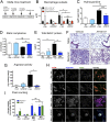
Macrophage depletion in MAFIA (MAcrophage Fas-Induced Apoptosis) mice results in decreased fibrosis. (A) Schematic showing the AP20187 treatment regimen that was used to deplete macrophages after bleomycin instillation in MAFIA mice. (B) Total macrophage numbers determined by flow cytometry from whole lung digest from bleomycin-instilled mice showed a significant decrease in the CD11c+CD11bvarF4/80+ and CD11cCD11b+F4/80+ subsets after delivery of AP20187 (*P < 0.05). There was no significant depletion of alveolar macrophages (P = 0.868). (C) Hydroxyproline levels were significantly reduced in bleomycin-instilled mice after delivery of AP20187 (***P < 0.001) compared with mice receiving bleomycin + vehicle. (D) Static compliance was significantly improved in AP20187-treated bleomycin-instilled MAFIA mice when compared with bleomycin-instilled mice receiving vehicle alone (**P < 0.01). (E) Total BALF protein levels were significantly reduced (**P < 0.01) in AP20187-treated bleomycin-instilled MAFIA mice compared with vehicle-treated bleomycin-instilled MAFIA mice. (F) Reduced lung collagen in Masson’s trichrome–stained lung sections from AP20187-treated bleomycin-instilled MAFIA mice compared with vehicle-treated bleomycin-instilled MAFIA mice. Original magnification: ×20. (G) Arginase activity in whole lung homogenates was significantly reduced (*P < 0.05) in AP20187-treated bleomycin-instilled MAFIA mice compared with vehicle-treated bleomycin-instilled MAFIA mice. (H) Immunofluorescent staining for F4/80 (red), arginase I (blue), and NOS2 (green) revealed that the remaining alveolar macrophages in AP20187-treated, bleomycin-instilled MAFIA mice exhibit a unique intermediate programming state in which both markers were expressed. (I) Quantitative fluorescence analysis by pixel counting showed that there was no significant difference (P = 0.7408) in the percentage of arginase I–positive macrophages between AP20187 and vehicle-treated, bleomycin-instilled MAFIA mice. There was a significant increase in the percentage of NOS2-staining macrophages (***P < 0.001).

References

    1. American Thoracic Society. Idiopathic pulmonary fibrosis: diagnosis and treatment. International consensus statement. American Thoracic Society (ATS), and the European Respiratory Society (ERS) Am J Respir Crit Care Med. 2000;161:646–664. - PubMed
    1. Raghu G, Weycker D, Edelsberg J, Bradford WZ, Oster G. Incidence and prevalence of idiopathic pulmonary fibrosis. Am J Respir Crit Care Med. 2006;174:810–816. - PubMed
    1. Myers JL, Katzenstein AL. Epithelial necrosis and alveolar collapse in the pathogenesis of usual interstitial pneumonia. Chest. 1988;94:1309–1311. - PubMed
    1. Thannickal VJ, Horowitz JC. Evolving concepts of apoptosis in idiopathic pulmonary fibrosis. Proc Am Thorac Soc. 2006;3:350–356. - PMC - PubMed
    1. Broekelmann TJ, Limper AH, Colby TV, McDonald JA. Transforming growth factor beta 1 is present at sites of extracellular matrix gene expression in human pulmonary fibrosis. Proc Natl Acad Sci USA. 1991;88:6642–6646. - PMC - PubMed

Publication types

MeSH terms