

Comparison of transverse analysis between posteroanterior cephalogram and cone-beam computed tomography
- PMID: 24325622
- PMCID: PMC8650435
- DOI: 10.2319/072613-555.1
Comparison of transverse analysis between posteroanterior cephalogram and cone-beam computed tomography
Abstract
Objectives: To evaluate maxillary and mandibular alveolar and basal bone widths using cone-beam computed tomography (CBCT) and to verify the correlation between CBCT images and posteroanterior (PA) cephalograms.
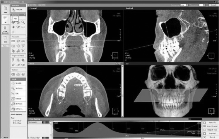
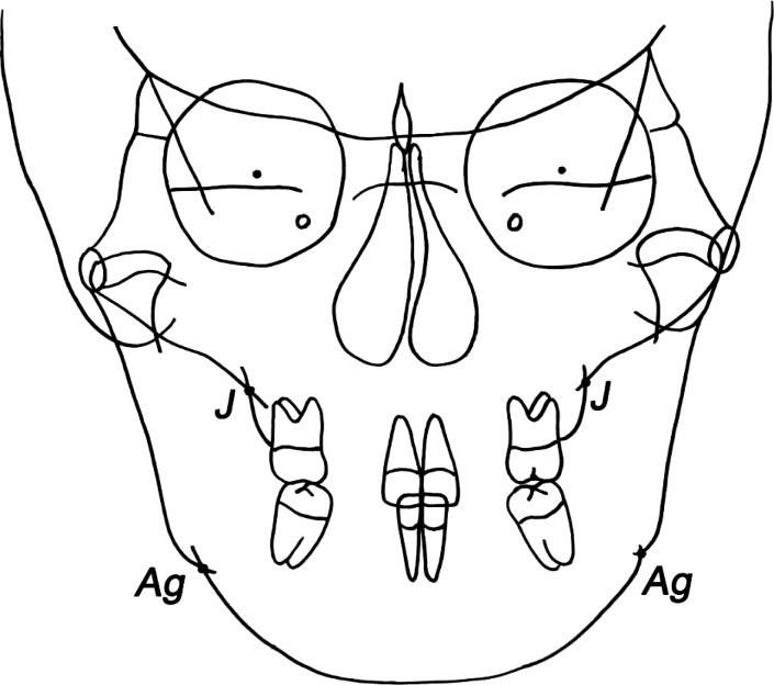
Materials and methods: The CBCT scans and PA cephalograms were obtained from 20 men (age range = 24.0-29.1 years; mean age = 27.2 years; SD = 2.8 years) and 20 women (age range = 20.3-28.1 years; mean age = 26.4 years; SD = 3.2 years) with normal occlusion. On CBCT images, maxillary and mandibular bone widths were measured at three posterior sites and five bone levels. The differences between maxillary and mandibular bone widths were calculated and compared with conventional transverse width of PA cephalograms.
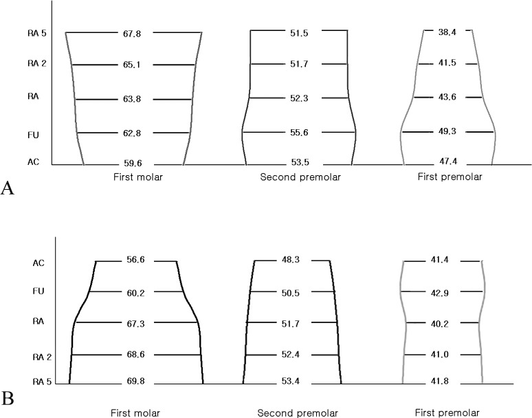
Results: Statistically significant differences in maxillary and mandibular bone widths were detected at different levels and sites. Bone widths were significantly increased from the alveolar crest toward the basal bone in the maxillary molar and mandibular second premolar and molar areas. A statistically significant correlation was only found between CBCT images and PA cephalograms for maxillomandibular width at the first molar area.
Conclusion: The results of this study suggested that three-dimensional assessment of maxillomandibular width is mandatory for the transverse analysis.
Keywords: CBCT, Transverse analysis; Maxillomandibular width.
Figures
References
-
- Vanarsdall RL, White RP., Jr Three-dimensional analysis for skeletal problems. Int J Adult Orthodon Orthognath Surg. 1994;9:159. - PubMed
-
- Vanarsdall RL., Jr Transverse dimension and long-term stability. Semin Orthod. 1999;5:171–180. - PubMed
-
- Betts NJ, Vanarsdall RL, Barber HD, Higgins-Barber K, Fonesca RJ. Diagnosis and treatment of transverse maxillary deficiency. Int J Adult Orthodon Orthognath Surg. 1995;10:75–96. - PubMed
-
- Ricketts RM. Perspectives in the clinical application of cephalometrics, the first fifty years. Angle Orthod. 1981;51:115–150. - PubMed
-
- Ghafari J, Cater PE, Shofer FS. Effect of film-object distance on posteroanterior cephalometric measurements: suggestions for standardized cephalometric methods. Am J Orthod Dentofacial Orthop. 1995;108:30–37. - PubMed
Publication types
MeSH terms
LinkOut - more resources
Full Text Sources
Other Literature Sources
