. 2013 Dec 10:10:149.
doi: 10.1186/1742-2094-10-149.

Minocycline prevents retinal inflammation and vascular permeability following ischemia-reperfusion injury

Affiliations

Minocycline prevents retinal inflammation and vascular permeability following ischemia-reperfusion injury

Steven F Abcouwer et al. J Neuroinflammation. .

Abstract

Background: Many retinal diseases are associated with vascular dysfunction accompanied by neuroinflammation. We examined the ability of minocycline (Mino), a tetracycline derivative with anti-inflammatory and neuroprotective properties, to prevent vascular permeability and inflammation following retinal ischemia-reperfusion (IR) injury, a model of retinal neurodegeneration with breakdown of the blood-retinal barrier (BRB).

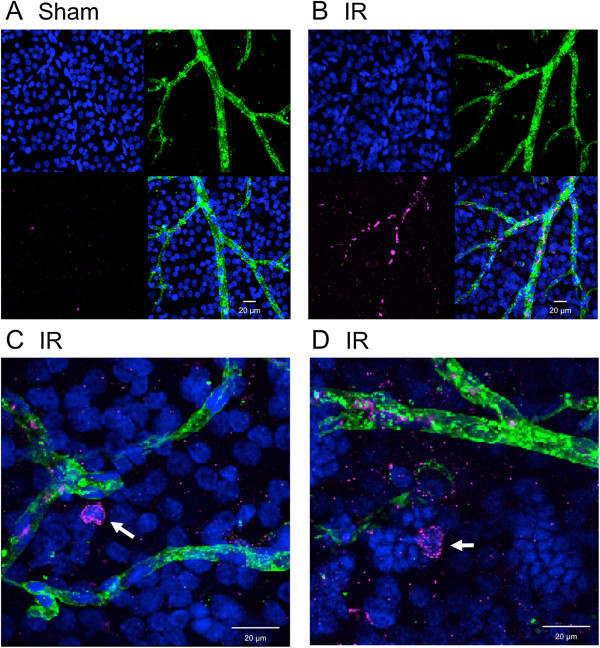
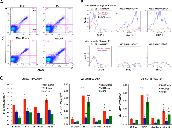
Methods: Male Sprague-Dawley rats were subjected to 45 min of pressure-induced retinal ischemia, with the contralateral eye serving as control. Rats were treated with Mino prior to and following IR. At 48 h after reperfusion, retinal gene expression, cellular inflammation, Evan's blue dye leakage, tight junction protein organization, caspase-3 activation, and DNA fragmentation were measured. Cellular inflammation was quantified by flow-cytometric evaluation of retinal tissue using the myeloid marker CD11b and leukocyte common antigen CD45 to differentiate and quantify CD11b+/CD45low microglia, CD11b+/CD45hi myeloid leukocytes and CD11bneg/CD45hi lymphocytes. Major histocompatibility complex class II (MHCII) immunoreactivity was used to determine the inflammatory state of these cells.

Results: Mino treatment significantly inhibited IR-induced retinal vascular permeability and disruption of tight junction organization. Retinal IR injury significantly altered mRNA expression for 21 of 25 inflammation- and gliosis-related genes examined. Of these, Mino treatment effectively attenuated IR-induced expression of lipocalin 2 (LCN2), serpin peptidase inhibitor clade A member 3 N (SERPINA3N), TNF receptor superfamily member 12A (TNFRSF12A), monocyte chemoattractant-1 (MCP-1, CCL2) and intercellular adhesion molecule-1 (ICAM-1). A marked increase in leukostasis of both myeloid leukocytes and lymphocytes was observed following IR. Mino treatment significantly reduced retinal leukocyte numbers following IR and was particularly effective in decreasing the appearance of MHCII+ inflammatory leukocytes. Surprisingly, Mino did not significantly inhibit retinal cell death in this model.

Conclusions: IR induces a retinal neuroinflammation within hours of reperfusion characterized by inflammatory gene expression, leukocyte adhesion and invasion, and vascular permeability. Despite Mino significantly inhibiting these responses, it failed to block neurodegeneration.


Figures

Figure 1
Figure 1
Minocycline (Mino) treatment significantly inhibited retinal vascular leakage and tight junction reorganization following ischemia-reperfusion (IR). Mino was delivered as twice-daily intraperitoneal (IP) injections, with two initial dosages of 45 mg/kg prior to ischemia and dosages of 22.5 mg/kg just prior to ischemia and every 12 h for the next 2 d during the reperfusion period as described in Materials and Methods. Non-treated (NT) animals received PBS vehicle injections. One eye of each rat was subjected to IR for 45 min or needle puncture only (Sham) and after 48 h of reperfusion retinas were assayed for (A) Evans blue dye leakage (n = 8 retinas per group), or (B and C) for vascular endothelium tight junction organization by immunohistochemistry of ZO-1 protein at endothelial cell borders (n = 4 retinas per group). Representative images from Sham and IR retinas are shown in B, with arrowheads indicating intact borders and arrows indicating regions of ZO-1 discontinuity. Distributions of endothelial cell border organization grading frequency are shown in C, with five representing fully continuous border staining and one representing complete loss of continuous border staining. *P ≤0.05, **P ≤0.01 and *** P ≤0.001 by Student’s t-test.
Figure 2
Figure 2
Minocycline (Mino) treatment significantly diminished neuroinflammatory gene expression following ischemia-reperfusion (IR) without affecting astrogliosis-related gene expression. Mino was delivered as twice-daily intraperitoneal (IP) injections, with two initial dosages of 45 mg/kg prior to ischemia and dosages of 22.5 mg/kg just prior to ischemia and every 12 h for the next 2 d during the reperfusion period as described in Materials and Methods. Non-treated (NT) animals received PBS vehicle injections. One eye of each rat was subjected to IR for 45 min or needle puncture only (Sham) and after 48 h of reperfusion total RNA was isolated from retinas and relative mRNA levels of neuroinflammation-related genes (A) ICAM-1, (B) LCN2, (C) TNFRSR12A, (D) SERPINA3N, (E) CCL2 and (F) the astrogliosis-related gene GFAP by duplex qRT-PCR with β-actin mRNA serving as control. Results shown are the means and standard error of means obtained from eight animals per group. *P ≤0.05, **P ≤0.01 and ***P ≤0.001 by Students t-test.
Figure 3
Figure 3
Leukostasis and retinal tissue invasion of CD45-positive leukocytes following ischemia-reperfusion (IR). Eyes were subjected to IR for 45 min or needle puncture only (Sham) and after 48 h of reperfusion retinas were removed, stained with antibodies to CD45 (magenta), isolectin B4 (IB4, green) and Hoechst nuclear stain (blue) and then flat mounted. Representative images showing CD45-staining leukocytes within IB4-stained vessels from IR retina (A) and sham retina (B) are shown along with magnified images (C and D) of CD45-positive cells (arrows) found within retinal tissue from IR retinas.
Figure 4
Figure 4
Minocycline (Mino) treatment significantly diminishes cellular inflammation following ischemia-reperfusion (IR). Mino was delivered as twice-daily intraperitoneal (IP) injections, with two initial dosages of 45 mg/kg prior to ischemia and dosages of 22.5 mg/kg just prior to ischemia and every 12 h for the next 2 d during the reperfusion period. Non-treated (NT) animals received PBS vehicle injections. One eye of each rat was subjected to IR for 45 min or needle puncture only (Sham) and after 48 h of reperfusion retinas were enzymatically dissociated and cells analyzed by flow cytometry. (A) Representative scatter plots of CD11b and CD45 immunostaining intensities of total retinal cells from Sham and IR retinas of rats treated with PBS or Mino. Events are partitioned into quadrant 1 (Q1) containing CD11b+/CD45low cells, quadrant 2 (Q2) containing CD11b+/CD45hi cells and quadrant 3 (Q3) containing CD11bneg/CD45hi cells. The fourth quadrant contains the majority of retinal cells, which are CD11bneg/CD45neg. (B) Example of MHCII staining distributions of cells from quadrants 1, 2 and 3. Red traces are for cells from Sham-treated retinas, blue traces are for cells from IR retinas and the black trace is for cells incubated without the MHCII antibody and used to define the intensity cutoff between define MHCII+ and MHCIIneg cells. (C) Quantification of total events, MHCII+ events and MHCIIneg events from quadrants 1, 2 and 3 from Sham and IR retinas from PBS- and Mino-treated rats. The numbers shown are mean percentiles relative to all events, including those in quadrant 4, obtained from three separate flow experiments, with a total of 11 to 12 determinations per group. Significant differences between Sham and IR groups are indicated as *P ≤0.05, **P ≤0.01 and ***P ≤0.001, while significant effects of Mino-treatment are indicated by #P ≤0.05 and ##P ≤0.01. All comparisons are by Students t-test.
Figure 5
Figure 5
Minocycline (Mino) treatment did not prevent cell death following ischemia-reperfusion (IR). (A and B) Mino was delivered as twice-daily intraperitoneal (IP) injections, with two initial dosages of 45 mg/kg prior to ischemia and dosages of 22.5 mg/kg just prior to ischemia and every 12 h for the next 2 d during the reperfusion period as described in Materials and Methods. One eye of each rat was subjected to IR for 45 min or needle puncture only (Sham) and after 48 h of reperfusion retinas were assayed for (A) caspase-3 activity and (B) DNA fragmentation cell death ELISA. (C) Mino dose–response. Rats were IP injected twice daily with PBS, 2.5 mg/kg (Mino-5), 7.5 mg/kg (Mino-15) or 22.5 mg/kg (Mino-45) beginning 24 h previous to ischemia, and 48 h following IR retinas were assayed by DNA fragmentation cell death ELISA. Results shown are the means and standard error of means obtained from eight animals per group. *P ≤0.05, **P ≤0.01 and ***P ≤0.001 by Student’s t-test.

References

    1. Osborne NN, Casson RJ, Wood JP, Chidlow G, Graham M, Melena J. Retinal ischemia: mechanisms of damage and potential therapeutic strategies. Prog Retin Eye Res. 2004;23:91–147. doi: 10.1016/j.preteyeres.2003.12.001. - DOI - PubMed
    1. Jehle T, Wingert K, Dimitriu C, Meschede W, Lasseck J, Bach M, Lagreze WA. Quantification of ischemic damage in the rat retina: a comparative study using evoked potentials, electroretinography, and histology. Invest Ophthalmol Vis Sci. 2008;49:1056–1064. doi: 10.1167/iovs.07-1050. - DOI - PubMed
    1. Husain S, Potter DE, Crosson CE. Opioid receptor-activation: retina protected from ischemic injury. Invest Ophthalmol Vis Sci. 2009;50:3853–3859. doi: 10.1167/iovs.08-2907. - DOI - PubMed
    1. Konno T, Sato A, Uchibori T, Nagai A, Kogi K, Nakahata N. Adenosine A2A receptor mediated protective effect of 2-(6-cyano-1-hexyn-1-yl)adenosine on retinal ischaemia/reperfusion damage in rats. Br J Ophthalmol. 2006;90:900–905. doi: 10.1136/bjo.2006.091496. - DOI - PMC - PubMed
    1. Nishijima K, Ng YS, Zhong L, Bradley J, Schubert W, Jo N, Akita J, Samuelsson SJ, Robinson GS, Adamis AP, Shima DT. Vascular endothelial growth factor-A is a survival factor for retinal neurons and a critical neuroprotectant during the adaptive response to ischemic injury. Am J Pathol. 2007;171:53–67. doi: 10.2353/ajpath.2007.061237. - DOI - PMC - PubMed

Publication types