. 2013;15(6):R212.
doi: 10.1186/ar4407.

Circulating levels of soluble MER in lupus reflect M2c activation of monocytes/macrophages, autoantibody specificities and disease activity

Circulating levels of soluble MER in lupus reflect M2c activation of monocytes/macrophages, autoantibody specificities and disease activity

Gaetano Zizzo et al. Arthritis Res Ther. 2013.

Abstract

Introduction: Systemic lupus erythematosus (SLE) is characterized by impaired efferocytosis and aberrant activation of innate immunity. We asked if shedding of MER receptor tyrosine kinase (MerTK) and AXL into soluble (s) ectodomains was related to immunological and clinical aspects of SLE.

Methods: Levels of sMER and sAXL in the plasma of 107 SLE patients and 45 matched controls were measured by ELISA. In 40 consecutive SLE patients, we examined potential correlations between either sMER or sAXL and plasma levels of sCD163, a marker of M2 activation. All three soluble receptors were measured in supernatants of monocytes/macrophages cultured in various immunological conditions. Membrane expression of MerTK, AXL and CD163 was assessed by flow cytometry.

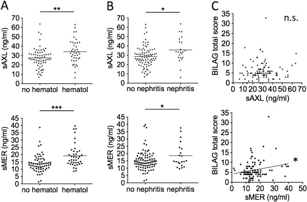
Results: Both sMER and sAXL were associated with anti-chromatin and anti-phospholipid autoantibodies, and with hematological and renal involvement. However, sMER and sAXL did not significantly correlate with each other; sAXL correlated with growth arrest-specific 6 (Gas6), whereas sMER correlated with reduced free protein S (PROS) levels. Only sMER showed significant associations with lupus-specific anti-dsDNA, anti-Sm, anti-ribonucleoprotein (anti-RNP) and anti-Ro60 autoantibodies. Strong correlations with disease activity indices (Systemic Lupus Erythematosus Disease Activity Index (SLEDAI), complement reduction, titer of circulating anti-dsDNA) were found for sMER, not for sAXL. Patients with active SLEDAI, nephritis, anti-dsDNA and anti-Ro60 positivity showed higher levels of sMER compared to controls. Levels of sMER, not sAXL, correlated with sCD163 levels, and these correlated with SLEDAI. Production of sMER and sCD163 occurred under “M2c” polarizing conditions, whereas sAXL was released upon type-I IFN exposure.

Conclusions: Alterations in homeostasis of anti-inflammatory and efferocytic “M2c” monocytes/macrophages may have a role in immunopathogenesis of SLE.


Figures

Figure 1
Figure 1
Plasma levels of soluble Mer are increased in discrete subsets of systemic lupus erythematosus patients. Levels of soluble Mer (sMer) or soluble Axl (sAxl) of 45 patients (pts) and 45 healthy controls (ctrls) were measured and compared by considering the whole groups (A) or by dividing patients and matched controls into subgroups according to disease activity (A), organ involvement (B) and autoantibody specificities (C, D and E). αCL, anticardiolipin immunoglobulin G; αDNA, anti-double-stranded DNA; αRo60, anti-Sjögren’s syndrome antigen A/Ro 60 KDa; SLEDAI, Systemic Lupus Erythematosus Disease Activity Index.
Figure 2
Figure 2
Soluble Axl and soluble Mer levels are associated with British Isles Lupus Assessment Group index hematological and renal involvement, and soluble Mer is also associated with total BILAG score. Levels of soluble Axl (sAxl) or soluble Mer (sMer) were measured in 107 matched and unmatched patients and compared according to active and/or stable (BILAG score ≥1) or inactive and/or absent (BILAG = 0) organ involvement (A) and (B). Associations with total BILAG score were then calculated (C). BILAG, British Isles Lupus Assessment Group index; hematol, hematological.*P < 0.05; **P < 0.01; ***P < 0.001; n.s., not significant.
Figure 3
Figure 3
Plasma levels of soluble Mer, but not soluble Axl, are closely associated with disease activity. Levels of soluble Axl (sAxl) and soluble Mer (sMer) were analyzed according to levels of complement fraction 3 (C3) (A) and C4 (B) and (C), circulating titers of anti-double-stranded DNA (anti-dsDNA) antibodies (D) and SLEDAI scores (E) and (F). SLEDAI, Systemic Lupus Erythematosus Disease Activity Index. *P < 0.05; **P < 0.01; ***P < 0.001; n.s., not significant.
Figure 4
Figure 4
Plasma levels of soluble Mer, but not soluble Axl, correlated with lupus autoantibody specificities. Levels of soluble Axl (sAxl) and soluble Mer (sMer) were analyzed according to the presence or absence of the following antibodies: antichromatin (αchrom) (A), anticardiolipin immunoglobulin G (IgG) (αCL) (B), lupus anticoagulant (LAC) (C), anti-double-stranded DNA (αdsDNA) (D), anti-Smith (αSm) (E), antiribonucleoprotein (αRNP) (F), anti-Sjögren’s syndrome antigen A (SSA)/Ro 52 and 60 KDa (αRo52 and αRo60) and anti-Sjögren’s syndrome antigen B (SSB)/La (G). *P < 0.05; **P < 0.01; ***P < 0.001.
Figure 5
Figure 5
Plasma levels of soluble Axl correlated with growth arrest–specific 6, but soluble Mer correlated with reduced free Protein S. Levels of soluble Axl (sAxl) and soluble Mer (sMer) were related to each other (A) and to Tyro3, Axl and MerTK (TAM) receptor ligands growth arrest–specific 6 (Gas6) and free Protein S (ProS) (B, C, D, and E). Cutoff values of Gas6 (ng/ml) and ProS (μg/ml) were established by considering mean values in patients and controls (Table 1). **P < 0.01; n.s., not significant.
Figure 6
Figure 6
Soluble Mer is released by M2c monocytes/macrophages and correlates with circulating levels of soluble CD163 in systemic lupus erythematosus, and soluble CD163, like soluble Mer, correlates with Systemic Lupus Erythematosus Disease Activity Index score. Levels of soluble Mer (sMer) (A) and soluble Axl (sAxl) (B) were measured in supernatants of monocytes/macrophages cultured for 3 days in the presence of medium alone (resting M0), interferon γ (IFN-γ), granulocyte macrophage colony-stimulating factor (GM-CSF), interleukin 17 (IL-17) (classically activated and/or proinflammatory M1 and M17), IL-4, transforming growth factor β (TGF-β), IL-10, macrophage colony-stimulating factor (M-CSF), dexamethasone (Dex), alone or in combination (alternatively activated and/or regulatory M2a and M2c cells). In 40 consecutive patients in our cohort, potential correlations between plasma levels of sMer (C) or sAxl (D) and soluble CD163 (sCD163) were examined. Levels of sCD163 were also analyzed according to circulating autoantibodies (E) and disease activity (F). αchrom., antichromatin; SLEDAI, Systemic Lupus Erythematosus Disease Activity Index. *P < 0.05; **P < 0.01; n.s., not significant.
Figure 7
Figure 7
Interferon α/β is required for production of soluble Axl, and combining M2c polarizing conditions with type I interferon exposure inhibits soluble Mer production and enhances soluble Axl release. Levels of soluble Mer (sMer) (A), soluble CD163 (sCD163) (B) and soluble Axl (sAxl) (C) were measured in supernatants of monocytes/macrophages cultured for 3 days in the presence or absence of type I interferons (IFN-α or IFN-β), macrophage growth factors (macrophage colony-stimulating factor (M-CSF) or granulocyte macrophage colony-stimulating factor (GM-CSF)), M2c polarizing agents (interleukin 10 (IL-10) or dexamethasone) or combinations of these. On day 2, low doses of lipopolysaccharide (LPS) were added for an additional 24 hours to stimulate ectodomain shedding of membrane receptors. Surface expression levels of Mer receptor tyrosine kinase (MerTK), Axl and CD163 were measured by flow cytometry (D). Numbers shown in (D) refer to mean fluorescence intensity values. Data are representative of three independent experiments. Dex, dexamethasone. *P < 0.05; **P < 0.01; ***P < 0.001; ****P < 0.0001.

References

    1. Herrmann M, Voll RE, Zoller OM, Hagenhofer M, Ponner BB, Kalden JR. Impaired phagocytosis of apoptotic cell material by monocyte-derived macrophages from patients with systemic lupus erythematosus. Arthritis Rheum. 1998;15:1241–1250. doi: 10.1002/1529-0131(199807)41:7<1241::AID-ART15>3.0.CO;2-H. - DOI - PubMed
    1. Muñoz LE, Lauber K, Schiller M, Manfredi AA, Herrmann M. The role of defective clearance of apoptotic cells in systemic autoimmunity. Nat Rev Rheumatol. 2010;15:280–289. doi: 10.1038/nrrheum.2010.46. - DOI - PubMed
    1. Shao WH, Cohen PL. Disturbances of apoptotic cell clearance in systemic lupus erythematosus. Arthritis Res Ther. 2011;15:202. doi: 10.1186/ar3535. - DOI - PMC - PubMed
    1. Wu X, Molinaro C, Johnson N, Casiano CA. Secondary necrosis is a source of proteolytically modified forms of specific intracellular autoantigens: implications for systemic autoimmunity. Arthritis Rheum. 2001;15:2642–2652. doi: 10.1002/1529-0131(200111)44:11<2642::AID-ART444>3.0.CO;2-8. - DOI - PubMed
    1. Vanden Berghe T, Vanlangenakker N, Parthoens E, Deckers W, Devos M, Festjens N, Guerin CJ, Brunk UT, Declercq W, Vandenabeele P. Necroptosis, necrosis and secondary necrosis converge on similar cellular disintegration features. Cell Death Differ. 2010;15:922–930. doi: 10.1038/cdd.2009.184. - DOI - PubMed

Publication types

MeSH terms