

Low-dose protocol of the spiral CT in orthodontics: comparative evaluation of entrance skin dose with traditional X-ray techniques
- PMID: 24325970
- PMCID: PMC4384968
- DOI: 10.1186/2196-1042-14-24
Low-dose protocol of the spiral CT in orthodontics: comparative evaluation of entrance skin dose with traditional X-ray techniques
Abstract
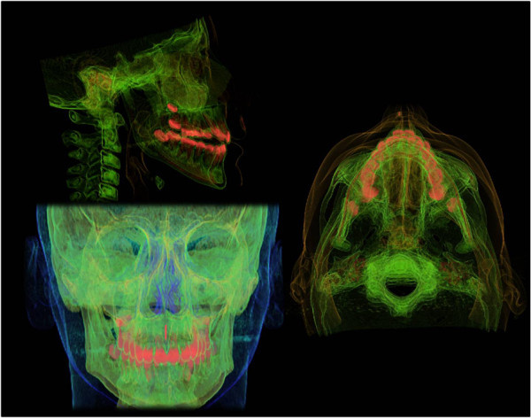
Background: The aim of this study was to evaluate the amount of radiation doses absorbed by soft tissues (entrance skin dose) with a low-dose spiral computed tomography (CT) protocol compared to conventional X-ray techniques commonly used in orthodontics.
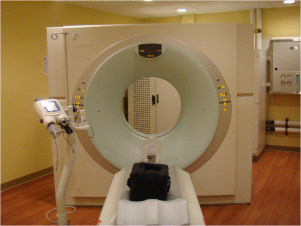
Methods: The amount of skin dose has been evaluated using a tissue-equivalent head-neck radiotherapy humanoid phantom with thermoluminescent dosimeters placed at the level of eye lens, parotid glands, and thyroid glands. CT images have been taken using a Sensation 16 Siemens CT scan and a low-dose protocol (15 mAs, 1 pitch, 2.5 mGy (CTDIvol), 80 kV, 1-mm slice thickness).
Results: The difference in image quality between traditional X-ray techniques and low-dose spiral CT was statistically significant (P<0.05). The difference in mean absorbed dose instead was not statistically significant.
Conclusions: Our protocol allows a more accurate orthodontic diagnosis without an increase of radiological risk for the patients in comparison to traditional X-ray techniques.
Figures
References
-
- Angle EH. Treatment of Malocclusion of the Teeth. Philadelphia: S.S. White; 1907.
-
- Moyers RE, Bookstein FL, Hunter WS. Analysis of the craniofacial skeleton: cephalometrics. In: Moyers RE, editor. Handbook of Orthodontics. 4. Chicago: Yearbook Medical Publishers; 1998. pp. 247–309.
Publication types
MeSH terms
LinkOut - more resources
Full Text Sources
Other Literature Sources
Medical
