. 2014 Jan 15;3(1):42-9.
doi: 10.1242/bio.20137120.

Mutagenesis and homologous recombination in Drosophila cell lines using CRISPR/Cas9

Affiliations

Mutagenesis and homologous recombination in Drosophila cell lines using CRISPR/Cas9

Andrew R Bassett et al. Biol Open. .

Abstract

We have applied the CRISPR/Cas9 system to Drosophila S2 cells to generate targeted genetic mutations in more than 85% of alleles. By targeting a constitutive exon of the AGO1 gene, we demonstrate homozygous mutation in up to 82% of cells, thereby allowing the study of genetic knockouts in a Drosophila cell line for the first time. We have shown that homologous gene targeting is possible at 1-4% efficiency using this system, allowing for the construction of defined insertions and deletions. We demonstrate that a 1 kb homology arm length is optimal for integration by homologous gene targeting, and demonstrate its efficacy by tagging the endogenous AGO1 protein. This technology enables controlled genetic manipulation in Drosophila cell lines, and its simplicity offers the opportunity to study cellular phenotypes genome-wide.

Keywords: CRISPR; Cas9; Drosophila S2 cells; Gene targeting; Genome engineering; Homologous recombination.


Conflict of interest statement

Competing interests: The authors have no competing interests to declare.

Figures

Fig. 1.
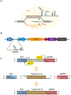
Fig. 1.. CRISPR/Cas9 expression system for Drosophila cell culture.
(A) The CRISPR/Cas9 system adapted from S. pyogenes for inducing double strand breaks. The synthetic guide RNA (sgRNA) contains 20 nt complementarity to the target site within the DNA, and the RNA structure necessary for incorporation into the Cas9 protein. Cas9 is indicated by a yellow circle, cleavage sites by arrowheads and protospacer adjacent motif (PAM, NGG) required for cleavage in red. (B) Schematic of the expression vector. The sgRNA is produced from a Drosophila U6 promoter by RNA polymerase III, which produces an uncapped transcript. Human codon-optimised Cas9 mRNA (orange oval) containing N- and C-terminal SV40 nuclear localisation signals (grey ovals) is produced from the strong, constitutive actin5C promoter by RNA polymerase II as the first half of a bicistronic transcript with the puromycin N-acetyltransferase gene (purple oval). The two open reading frames are separated by a viral 2A ribosome skipping site (red oval) to allow bicistronic expression, and transcription is terminated by a polyadenylation signal from the SV40 virus. (C) Strategy for cloning of target oligos. Two Bsp QI sites (yellow) cause cleavage at the end of the U6 promoter (blue) and sgRNA backbone (red), leaving 3 nt 5′ overhangs. Transcription from the dU6 promoter begins with the indicated G nucleotide (arrow). Target oligos (orange) are designed to provide complementary overhangs flanking the 20 nt target sequence. If the target sequence does not begin with a G, this is appended to its 5′ end to reconstitute the G nucleotide required by the dU6 promoter (as indicated here). See also supplementary material Table S1.
Fig. 2.
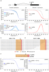
Fig. 2.. Mutagenesis of the yellow gene is highly efficient.
(A) Schematic of the yellow gene showing position of sgRNA target site. Exons are indicated as black boxes, transcriptional start site by an arrow and the y1 sgRNA target site by a black triangle. (B) Indel detection by high resolution melt analysis (HRMA) after transient transfection. Cells were transfected with empty vector lacking sgRNA (EV, grey lines) or with different amounts of vector expressing the y1 sgRNA (2, 1, 0.5 µg, blue lines). DNA was analysed by HRMA 3 days post-transfection, and indicated as melting curves (upper panel) or change relative to control (lower panel). (C) Sequencing of indel mutations after transient transfection. PCR products spanning the cleavage site (black triangle) were cloned and sequenced from cells transfected with 2 µg y1 sgRNA vector, and showed deletions in 2 of 18 clones sequenced at the expected site. Target site is highlighted in orange, and PAM in red. The first line of each alignment indicates the wt sequence. (D) As panel B for cells selected for a further 7 days in puromycin. (E) As panel C for cells selected for a further 7 days in puromycin. (F) Lack of off target mutagenesis at the CG14073 gene. Base pairing interactions between the y1 sgRNA and the site in CG14073 are indicated (target site indicated in orange, PAM in red, cleavage site as a black triangle). HRMA analysis at the closest off-target site in the Drosophila genome indicates a lack of detectable indel mutants even after 7 days selection in puromycin (as panels D,E). Primer sequences are indicated in supplementary material Table S1.
Fig. 3.
Fig. 3.. Homologous recombination at the yellow gene.
(A) Schematic diagram of the yellow gene. Exons are indicated by black boxes, y1 sgRNA target site as a black triangle, and position of homology arms by black boxes. Inserted sequence is indicated by a red box. PCR primer pairs used in Fig. 3B are indicated by arrows and a or b. Primer sequences are indicated in supplementary material Table S1 (aF, aR, bF, bR). (B) Integration of homology constructs at target site as detected by PCR. PCR was performed after transfection of cells with the indicated constructs to detect integration (upper panel, a) or as a control, outside of the integration site (lower panel, b). Cells were harvested 3 days after transfection (left, transient expression) or after a further 7 days selection in puromycin (right, selected 7 d puromycin). Integration was observed with all homology constructs, but only upon cotransfection with plasmid expressing both Cas9 and the y1 sgRNA. Efficiencies of correct integration are indicated (% targeting), and were normalised to control PCR product (lower panel, b). Primer sequences are indicated in supplementary material Table S1.
Fig. 4.
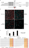
Fig. 4.. Mutagenesis of the AGO1 gene.
(A) Schematic of the AGO1 locus. Exons are indicated by boxes, coding sequence in black (CDS) and untranslated regions in grey (UTR). Different splice isoforms and an overlapping gene are indicated on different lines (AGO1-RA, RB, RC, RD, mRpL53). The position of the sgRNA target site is indicated by a black triangle, and the homology construct by a black rectangle, showing the inserted HA tag in red. Primer sequences are indicated in supplementary material Table S1. (B) Efficient mutagenesis of the AGO1 gene. Images show immunostaining with an anti-AGO1 antibody (red) counterstained for DNA to highlight nuclei of all cells (cyan). The upper left panel indicates cells transfected with Cas9 but lacking sgRNA (empty vector) analysed after selection for 7 days in puromycin. Other panels show cells transfected with the AGO1 sgRNA and analysed 3 days post transfection (upper right, transient expression), or selected for a further 3 or 7 days in puromycin (bottom panels, selected 3 d or 7 d puromycin). Cells are clearly visible that lack staining for AGO1 protein, but maintain nuclear staining in those cells transfected with the AGO1 sgRNA (examples indicated with white arrowheads). See also supplementary material Fig. S1. (C) Quantification of mutagenesis efficiency at the AGO1 gene. Cells that lacked visible staining for AGO1 protein were counted upon transfection with vector expressing AGO1 sgRNA (black), or lacking sgRNA expression (grey). Cells were analysed 3 days after transfection (transient) or after a further 3 or 7 days selection in puromycin (Selected 3 d or 7 d). Values were expressed as % mutant cells, and error bars indicate 95% confidence intervals of at least 4 biological repeats of at least 200 cells per repeat. (D) Sequencing of indel mutations within AGO1. PCR products spanning the cleavage site (black triangle) were cloned and sequenced from cells 3 d after transfection with 2 µg y1 sgRNA vector (Transient), or after a further 3 d selection in puromycin (Selected 3 d). Deletions in 9/23 (39.1%, Transient) or 21/26 (80.7%, Selected 3 d) clones were observed at the expected site. Target site is highlighted in orange, and PAM in red. The first line of each alignment indicates the wt sequence. Scale bars: 20 µm.
Fig. 5.
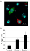
Fig. 5.. Epitope tagging of the AGO1 gene.
(A) Tagging of AGO1 with a HA epitope tag. Cells transfected with the AGO1 sgRNA and homology construct shown in Fig. 4A were analysed by immunostaining with anti-AGO1 (red) and anti-HA (green) antibodies and counterstained for DNA (cyan). Staining with anti-HA antibody is clearly observed in some cells (open arrowhead), with a cytoplasmic distribution similar to that of AGO1. Many cells show no AGO1 staining (closed arrowhead) due to homozygous mutation of the endogenous AGO1 gene. (B) Quantification of AGO1 tagging efficiency. Number of cells staining with anti-HA antibody were quantified 3 days after transfection (transient), or after a further 4 or 7 days selection in puromycin (Selected 4 d or 7 d). Values were expressed as a percentage of the total number of cells (AGO1 wt and mutant), and error bars indicate 95% confidence intervals of at least 4 biological repeats of at least 200 cells per repeat. Scale bar: 10 µm.

References

    1. Barrangou R., Fremaux C., Deveau H., Richards M., Boyaval P., Moineau S., Romero D. A., Horvath P. (2007). CRISPR provides acquired resistance against viruses in prokaryotes. Science 315, 1709–1712 10.1126/science.1138140 - DOI - PubMed
    1. Bassett A. R., Tibbit C., Ponting C. P., Liu J. L. (2013). Highly efficient targeted mutagenesis of Drosophila with the CRISPR/Cas9 system. Cell Rep 4, 220–228 10.1016/j.celrep.2013.06.020 - DOI - PMC - PubMed
    1. Bedell V. M., Wang Y., Campbell J. M., Poshusta T. L., Starker C. G., Krug R. G. I. I., Tan W., Penheiter S. G., Ma A. C., Leung A. Y. et al. (2012). In vivo genome editing using a high-efficiency TALEN system. Nature 491, 114–118 10.1038/nature11537 - DOI - PMC - PubMed
    1. Beumer K., Bhattacharyya G., Bibikova M., Trautman J. K., Carroll D. (2006). Efficient gene targeting in Drosophila with zinc-finger nucleases. Genetics 172, 2391–2403 10.1534/genetics.105.052829 - DOI - PMC - PubMed
    1. Beumer K. J., Trautman J. K., Bozas A., Liu J. L., Rutter J., Gall J. G., Carroll D. (2008). Efficient gene targeting in Drosophila by direct embryo injection with zinc-finger nucleases. Proc. Natl. Acad. Sci. USA 105, 19821–19826 10.1073/pnas.0810475105 - DOI - PMC - PubMed