Comparative Study
. 2014 Mar;34(3):450-9.
doi: 10.1038/jcbfm.2013.217. Epub 2013 Dec 11.

Immune cell infiltration in malignant middle cerebral artery infarction: comparison with transient cerebral ischemia

Affiliations
Comparative Study

Immune cell infiltration in malignant middle cerebral artery infarction: comparison with transient cerebral ischemia

Hannah X Chu et al. J Cereb Blood Flow Metab. 2014 Mar.

Abstract

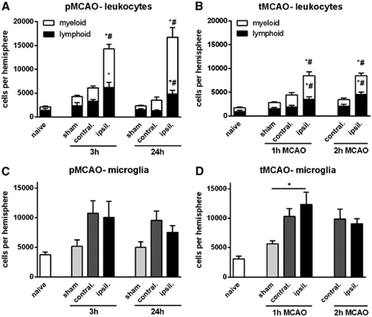
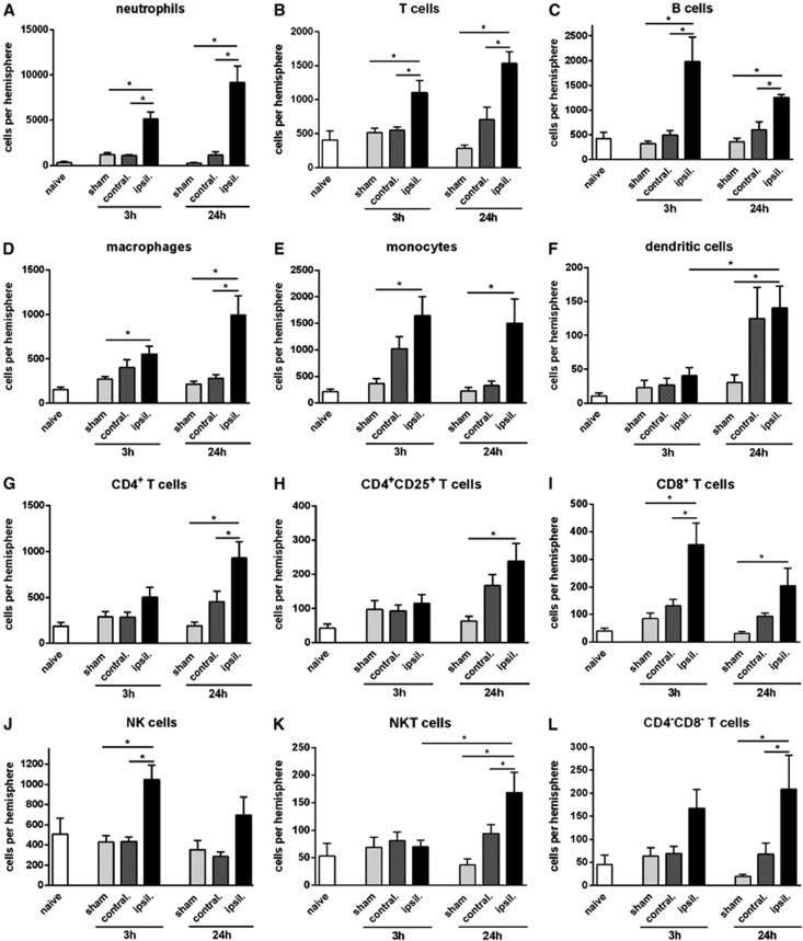
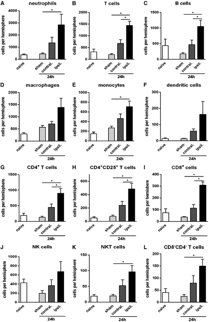
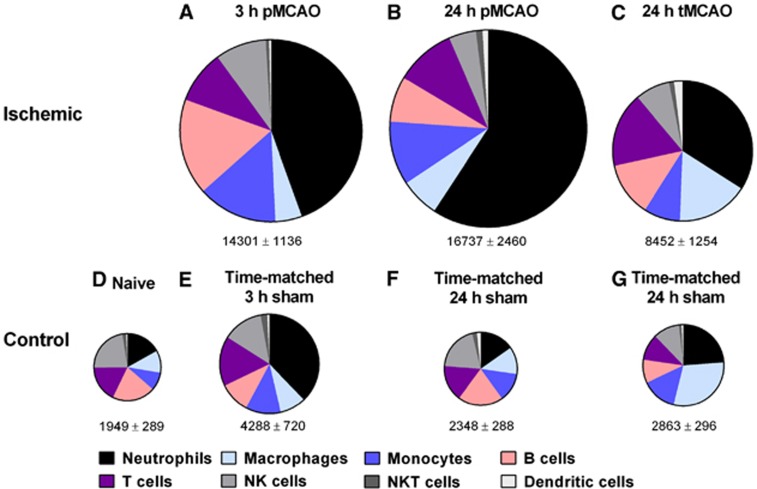
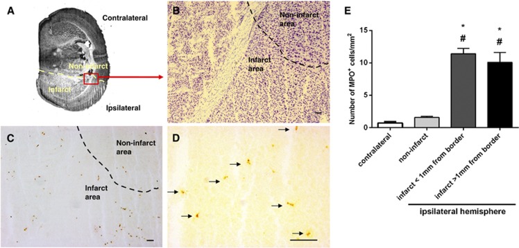
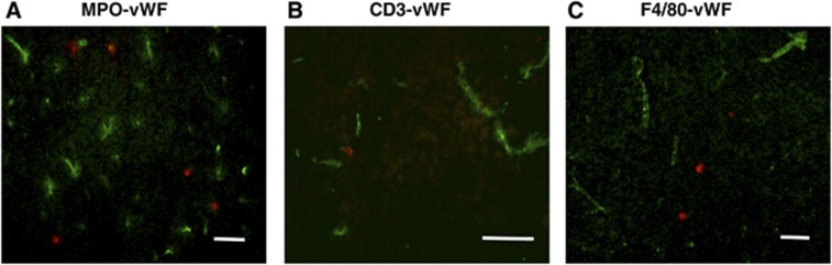
We tested whether significant leukocyte infiltration occurs in a mouse model of permanent cerebral ischemia. C57BL6/J male mice underwent either permanent (3 or 24 hours) or transient (1 or 2 hours+22- to 23-hour reperfusion) middle cerebral artery occlusion (MCAO). Using flow cytometry, we observed ∼15,000 leukocytes (CD45(+high) cells) in the ischemic hemisphere as early as 3 hours after permanent MCAO (pMCAO), comprising ∼40% lymphoid cells and ∼60% myeloid cells. Neutrophils were the predominant cell type entering the brain, and were increased to ∼5,000 as early as 3 hours after pMCAO. Several cell types (monocytes, macrophages, B lymphocytes, CD8(+) T lymphocytes, and natural killer cells) were also increased at 3 hours to levels sustained for 24 hours, whereas others (CD4(+) T cells, natural killer T cells, and dendritic cells) were unchanged at 3 hours, but were increased by 24 hours after pMCAO. Immunohistochemical analysis revealed that leukocytes typically had entered and widely dispersed throughout the parenchyma of the infarct within 3 hours. Moreover, compared with pMCAO, there were ∼50% fewer infiltrating leukocytes at 24 hours after transient MCAO (tMCAO), independent of infarct size. Microglial cell numbers were bilaterally increased in both models. These findings indicate that a profound infiltration of inflammatory cells occurs in the brain early after focal ischemia, especially without reperfusion.


Figures

Figure 1
Figure 1
Flow cytometric quantification of leukocytes in the brain, expressed as total, myeloid, or lymphoid cells, at 3 or 24 hours after permanent middle cerebral artery occlusion (pMCAO) (A), or at 24 hours after 1- or 2-hour transient middle cerebral artery occlusion (tMCAO) (B). Data are shown for the contralateral and ischemic hemispheres, and compared with data from naïve or time-matched sham control animals. (n=5 to 10; *P<0.05 versus sham; #P<0.05 versus contralateral; one-way analysis of variance (ANOVA) with Bonferroni post tests). Microglia numbers are also shown after pMCAO (C) and tMCAO (D) (n=5 to 10; *P<0.05 versus sham; #P<0.05 versus contralateral; one-way ANOVA with Bonferroni post test). Data are mean±s.e.m.
Figure 2
Figure 2
Quantification of leukocyte subtypes in brain at 3 or 24 hours after permanent middle cerebral artery occlusion, compared with naïve and time-matched sham controls (n=5 to 10, *P<0.05; one-way analysis of variance (ANOVA) with Bonferroni post tests). Data are shown for neutrophils (A), T cells (B), B cells (C), macrophages (D), monocytes (E), dendritic cells (F), CD4+ T cells (G), CD4+CD25+ T cells (H), CD8+ T cells (I), natural killer (NK) cells (J), natural killer T (NKT) cells (K), and CD4CD8 T cells (L), and expressed as mean±s.e.m.
Figure 3
Figure 3
Quantification of leukocyte subtypes in brain at 24 hours after transient middle cerebral artery occlusion, compared with naïve and time-matched sham controls (n=5, *P<0.05; one-way analysis of variance (ANOVA) with Bonferroni post tests). Data are shown for neutrophils (A), T cells (B), B cells (C), macrophages (D), monocytes (E), dendritic cells (F), CD4+ T cells (G), CD4+CD25+ T cells (H), CD8+ T cells (I), natural killer (NK) cells (J), natural killer T (NKT) cells (K), and CD4CD8 T cells (L), and expressed as mean±s.e.m.
Figure 4
Figure 4
Pie charts summarizing number and composition of immune cells in ischemic hemispheres of mouse brains at 3 hours (A) or 24 hours (B) after permanent middle cerebral artery occlusion (pMCAO) or at 24 hours after 1 hour of transient middle cerebral artery occlusion (tMCAO) (C), and of brain hemispheres from naïve (D) or time-matched sham (EG) control mice. The area of each pie is proportional to the number of leukocytes per hemisphere (also shown as mean±s.e.m.). n=20 for naïve; n=10 to 15 for all other groups.
Figure 5
Figure 5
Representative photomicrographs showing localization of myeloperoxidase (MPO+) cells in mouse brain at 3 hour after permanent middle cerebral artery occlusion (pMCAO). (A) Low magnification of a thionin-stained section that outlines the infarct area (lighter in color; border shown as dashed line). (B) The perilesional area from the ischemic hemisphere of a thionin-stained coronal brain section, as observed under × 100 magnification. (C) A consecutive section was stained with anti-MPO antibody and examined under the same magnification; brown spots indicate MPO+ cells. The majority of MPO+ cells were present within the infarct. (D) Granule-containing MPO+ cells (indicated by arrows) were observed under further magnification ( × 400). Scale bars in (BD)=100 μm. (E) Quantification of MPO+ cell density in the contralateral hemisphere, noninfarcted areas of the ischemic hemisphere, and infarcted areas (either within 1 mm of the infarct border or deeper in the infarct core >1 mm from infarct border). *P<0.05 compared with contralateral hemisphere; #P<0.05 compared with noninfarcted area. n=a total of 128 high power fields of brain sections from 4 mice, one-way analysis of variance (ANOVA), with Bonferroni post tests. Data are mean±s.e.m.
Figure 6
Figure 6
Neutrophils, T cells, and macrophages are infiltrated into the ischemic infarct at 3 hours after permanent middle cerebral artery occlusion (pMCAO). Representative overlayed images of myeloperoxidase (MPO+) cells (A), CD3+ cells (B), or F4/80+ cells (C) (red fluorescence) with endothelial marker von Willebrand Factor (vWF; green fluorescence) show that most immune cells are located outside the vasculature. Scale bar=50 μm.

References

    1. Roger VL, Go AS, Lloyd-Jones DM, Benjamin EJ, Berry JD, Borden WB, et al. Heart disease and stroke statistics—2012 update: a report from the American Heart Association. Circulation. 2012;125:e2–e220. - PMC - PubMed
    1. Del Zoppo GJ, Saver JL, Jauch EC, Adams HP., Jr. Expansion of the time window for treatment of acute ischemic stroke with intravenous tissue plasminogen activator: a science advisory from the American Heart Association/American Stroke Association. Stroke. 2009;40:2945–2948. - PMC - PubMed
    1. Frank JI. Large hemispheric infarction, deterioration, and intracranial pressure. Neurology. 1995;45:1286–1290. - PubMed
    1. Berrouschot J, Sterker M, Bettin S, Koster J, Schneider D. Mortality of space-occupying ('malignant') middle cerebral artery infarction under conservative intensive care. Intensive Care Med. 1998;24:620–623. - PubMed
    1. Hacke W, Schwab S, Horn M, Spranger M, De Georgia M, von Kummer R. 'Malignant' middle cerebral artery territory infarction: clinical course and prognostic signs. Arch Neurol. 1996;53:309–315. - PubMed

Publication types