. 2013 Dec 10;4(6):e00707-13.
doi: 10.1128/mBio.00707-13.

The intestinal microbiota interferes with the microRNA response upon oral Listeria infection

Affiliations

The intestinal microbiota interferes with the microRNA response upon oral Listeria infection

Cristel Archambaud et al. mBio. .

Abstract

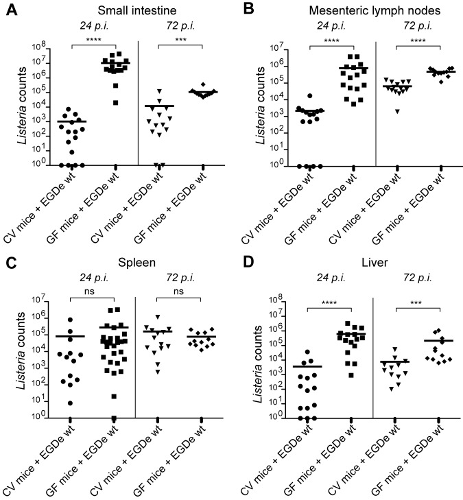
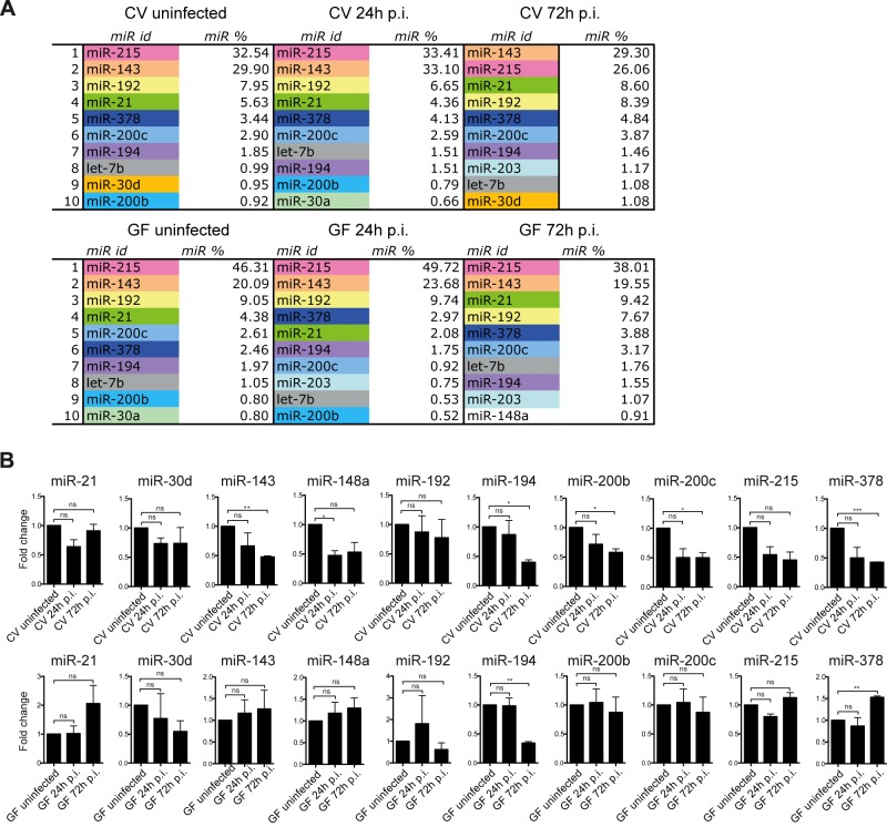
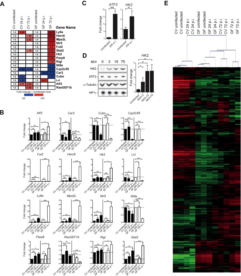
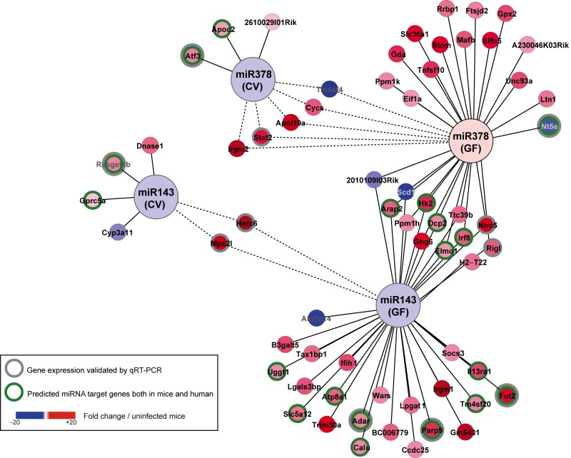
The intestinal tract is the largest reservoir of microbes in the human body. The intestinal microbiota is thought to be able to modulate alterations of the gut induced by enteropathogens, thereby maintaining homeostasis. Listeria monocytogenes is the agent of listeriosis, an infection transmitted to humans upon ingestion of contaminated food. Crossing of the intestinal barrier is a critical step of the infection before dissemination into deeper organs. Here, we investigated the role of the intestinal microbiota in the regulation of host protein-coding genes and microRNA (miRNA or miR) expression during Listeria infection. We first established the intestinal miRNA signatures corresponding to the 10 most highly expressed miRNAs in the murine ileum of conventional and germfree mice, noninfected and infected with Listeria. Next, we identified 6 miRNAs whose expression decreased upon Listeria infection in conventional mice. Strikingly, five of these miRNA expression variations (in miR-143, miR-148a, miR-200b, miR-200c, and miR-378) were dependent on the presence of the microbiota. In addition, as is already known, protein-coding genes were highly affected by infection in both conventional and germfree mice. By crossing bioinformatically the predicted targets of the miRNAs to our whole-genome transcriptomic data, we revealed an miRNA-mRNA network that suggested miRNA-mediated global regulation during intestinal infection. Other recent studies have revealed an miRNA response to either bacterial pathogens or commensal bacteria. In contrast, our work provides an unprecedented insight into the impact of the intestinal microbiota on host transcriptional reprogramming during infection by a human pathogen.

Importance: While the crucial role of miRNAs in regulating the host response to bacterial infection is increasingly recognized, the involvement of the intestinal microbiota in the regulation of miRNA expression has not been explored in detail. Here, we investigated the impact of the intestinal microbiota on the regulation of protein-coding genes and miRNA expression in a host infected by L. monocytogenes, a food-borne pathogen. We show that the microbiota interferes with the microRNA response upon oral Listeria infection and identify several protein-coding target genes whose expression correlates inversely with that of the miRNA. Further investigations of the regulatory networks involving miR-143, miR-148a, miR-200b, miR-200c, and miR-378 will provide new insights into the impact of the intestinal microbiota on the host upon bacterial infection.


Figures

FIG 1
FIG 1
Germfree mice are hypersensitive to Listeria infection. Listeria counts were assessed in the small intestine (A), mesenteric lymph nodes (B), spleen (C), and liver (D) of conventional (CV) and germfree (GF) wild-type mice uninfected or orally infected with L. monocytogenes EGD-e wild-type (EGDe wt) for 24 h and 72 h (24 h p.i. and 72 h p.i.). Each dot represents one organ. Horizontal bars represent the mean for each condition. Statistical tests were performed using a Mann-Whitney test. Asterisks indicate a P value considered statistically significant (***, P < 0.001; ****, P < 0.0001); ns, nonsignificant difference.
FIG 2
FIG 2
Listeria and the intestinal microbiota slightly affect the intestinal miRNA expression pattern. (A) The 10 most highly expressed miRNAs (miR) in the small intestine of conventional (CV) and germfree (GF) mice uninfected or orally infected with L. monocytogenes for 24 h and 72 h (24 h p.i. and 72 h p.i.) were determined. In each list, the identity of each individual miR (miR id) is indicated, as well as its percentage (miR %) of the total miRs. (B) Relative expression levels of miR-21, miR-30d, miR-143, miR-148a, miR-192, miR-194, miR-200b, miR-200c, miR-215, and miR-378 in conventional (CV) and germfree (GF) mice orally infected with L. monocytogenes for 24 h and 72 h (24 h p.i. and 72 h p.i.). Shown are fold changes after standardization to the small nuclear RNA U6 and using uninfected CV and GF control mice as references. Data are represented as means with standard errors of the means (SEM) of values for individual mice (n ≥ 3 per group) from three independent experiments. Statistical tests were performed using a two-tailed Student’s t test. Asterisks indicate a value considered statistically significant (*, P < 0.05; **, P < 0.01; ***, P < 0.001); ns, nonsignificant difference.
FIG 3
FIG 3
The intestinal microbiota affects the transcriptional host response to Listeria infection. (A) The heat map presents a subset of 16 representative host genes whose expression was significantly affected (false discovery rate, Benjamini and Hochberg approach [FDR-BH], P < 0.05) in the small intestine of conventional (CV) and germfree (GF) mice (n = 2) uninfected or orally infected with L. monocytogenes for 24 h and 72 h (24 h p.i. and 72 h p.i.). The first three columns show the fold changes of gene expression levels in CV mice at 24 h p.i. and 72 h p.i. relative to the expression levels in uninfected CV mice. The last three columns show the fold changes of gene expression levels in GF mice at 24 h p.i. and 72 h p.i. relative to the expression levels in uninfected GF mice. White squares indicate genes whose expression was not significantly affected by the Listeria infection. (B) Relative expression levels of the 16 host genes in CV and GF mice orally infected with L. monocytogenes 24 h p.i. and 72 h p.i. Shown are fold changes after standardization to GADPH and using uninfected CV and GF control mice as references. Data are represented as means and SEM of values for individual mice (n ≥ 3 per group) from three independent experiments. (C) Relative abundance of ATF3 and HK2 transcripts in LoVo cells infected with L. monocytogenes for 20 h. Shown are fold changes after standardization to the GAPDH transcript and using uninfected LoVo cells as a reference. Error bars indicate standard deviations. (D) Relative levels of abundance of ATF3 and HK2 proteins in LoVo cells infected with L. monocytogenes for 22 h shown by Western blot analysis of cell extracts of uninfected cells or cells infected at an MOI of 3, 15, or 75 (left). Quantification of the Western blot signals for the HK2 protein (right). Shown are fold changes after standardization to the α-tubulin protein and using uninfected LoVo cells as a reference. Error bars indicate standard deviations. (E) CV and GF mice were orally infected with L. monocytogenes for 24 h p.i. and 72 h p.i. (2 mice per condition). As described in the text, 444 genes were identified as displaying significantly changed expression levels under at least one of the 6 tested conditions. Unsupervised hierarchical clustering based on this list of the 444 genes was performed with the MeV application. The red–green color shows log2 ratios from mean centered gene expression levels and indicates an upregulation and a downregulation compared to the mean, respectively. See Table S1 in the supplemental material for a list of the 444 genes and their corresponding expression values. Statistics were performed using a two-tailed Student’s t test. Asterisks indicate a value considered statistically significant (*, P < 0.05; **, P < 0.01; ***, P < 0.001; ****, P < 0.0001); ns, nonsignificant difference.
FIG 4
FIG 4
miRNA-mRNA network. Using Cytoscape, we highlighted, among the 5 miRNAs whose expression was affected by L. monocytogenes and the intestinal microbiota, a network linking miR-143 and miR-378 and the regulated host genes in the small intestine of conventional (CV) and germfree (GF) mice orally infected with L. monocytogenes 72 h p.i. As described in the text, miR-143 decreased upon infection in both CV and GF mice; in contrast, miR-378 decreased in CV mice but not in GF mice. Nodes represent infection-regulated miRNAs and genes; lines link each miRNA to its putative targets. Some of the predictions in mice were valid in humans (green circles). The color coding indicates the fold change of gene expression in Listeria-infected mice relative to the expression level in uninfected control mice. The differential expression levels of 11 genes were validated by RT-qPCR, as shown in Fig. 3B. Dotted lines show a gene that is similarly regulated in CV and GF mice.

References

    1. Cossart P. 2011. Illuminating the landscape of host-pathogen interactions with the bacterium Listeria monocytogenes. Proc. Natl. Acad. Sci. U. S. A. 108:19484–19491 - PMC - PubMed
    1. Stavru F, Archambaud C, Cossart P. 2011. Cell biology and immunology of Listeria monocytogenes infections: novel insights. Immunol. Rev. 240:160–184 - PubMed
    1. Aubry C, Goulard C, Nahori MA, Cayet N, Decalf J, Sachse M, Boneca IG, Cossart P, Dussurget O. 2011. OatA, a peptidoglycan O-acetyltransferase involved in Listeria monocytogenes immune escape, is critical for virulence. J. Infect. Dis. 204:731–740 - PMC - PubMed
    1. Boneca IG, Dussurget O, Cabanes D, Nahori MA, Sousa S, Lecuit M, Psylinakis E, Bouriotis V, Hugot JP, Giovannini M, Coyle A, Bertin J, Namane A, Rousselle JC, Cayet N, Prévost MC, Balloy V, Chignard M, Philpott DJ, Cossart P, Girardin SE. 2007. A critical role for peptidoglycan N-deacetylation in Listeria evasion from the host innate immune system. Proc. Natl. Acad. Sci. U. S. A. 104:997–1002 - PMC - PubMed
    1. Rae CS, Geissler A, Adamson PC, Portnoy DA. 2011. Mutations of the Listeria monocytogenes peptidoglycan N-deacetylase and O-acetylase result in enhanced lysozyme sensitivity, bacteriolysis, and hyperinduction of innate immune pathways. Infect. Immun. 79:3596–3606 - PMC - PubMed

Publication types