The ocular surface phenotype of Muc5ac and Muc5b null mice
- PMID: 24327612
- PMCID: PMC3894795
- DOI: 10.1167/iovs.13-13194
The ocular surface phenotype of Muc5ac and Muc5b null mice
Abstract
Purpose: Recent development of mice null for either Muc5ac or Muc5b mucin allows study of their specific roles at the mouse ocular surface. A recent report indicated that Muc5ac null mice show an ocular surface phenotype similar to that seen in dry eye syndrome. The purpose of our study was to determine the effect of lack of Muc5ac or Muc5b on the ocular surface, and to determine if environmental desiccating stress exacerbated a phenotype.
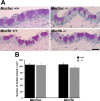
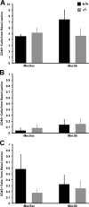
Methods: Muc5ac null and Muc5b null mice, and their wild-type controls were examined for ocular surface defects by fluorescein staining. The number of goblet cells per area of conjunctival epithelium was counted, and levels of mucin gene expression and genes associated with epithelial stress, keratinization, and differentiation, known to be altered in dry eye syndrome, were assayed. To determine if the null mice would respond more to desiccating stress than their wild-type controls, they were challenged in a controlled environment chamber (CEC) and assessed for changes in fluorescein staining, tear volume, and inflammatory cells within the conjunctival and corneal epithelia.
Results: Unlike the previous study, we found no ocular surface phenotype in the Muc5ac null mice, even after exposure to desiccating environmental stress. Similarly, no ocular surface phenotype was present in the Muc5b null mice, either before or after exposure to a dry environment in the CEC.
Conclusions: Our results indicate that deleting either the Muc5ac or Muc5b gene is insufficient to create an observable dry eye phenotype on the ocular surface of these mice.
Keywords: animal models; dry eye; mucins.
Figures


Similar articles
-
Entrapment of conjunctival goblet cells by desiccation-induced cornification.Invest Ophthalmol Vis Sci. 2011 Jun 1;52(6):3492-9. doi: 10.1167/iovs.10-5782. Invest Ophthalmol Vis Sci. 2011. PMID: 21421863 Free PMC article.
-
Spdef null mice lack conjunctival goblet cells and provide a model of dry eye.Am J Pathol. 2013 Jul;183(1):35-48. doi: 10.1016/j.ajpath.2013.03.017. Epub 2013 May 10. Am J Pathol. 2013. PMID: 23665202 Free PMC article.
-
A nanomedicine to treat ocular surface inflammation: performance on an experimental dry eye murine model.Gene Ther. 2013 May;20(5):467-77. doi: 10.1038/gt.2012.56. Epub 2012 Jul 19. Gene Ther. 2013. PMID: 22809996
-
Secreted Mucins on the Ocular Surface.Invest Ophthalmol Vis Sci. 2018 Nov 1;59(14):DES151-DES156. doi: 10.1167/iovs.17-23623. Invest Ophthalmol Vis Sci. 2018. PMID: 30481820 Review.
-
Significance of mucin on the ocular surface.Cornea. 2002 Mar;21(2 Suppl 1):S17-22. doi: 10.1097/00003226-200203001-00005. Cornea. 2002. PMID: 11995804 Review.
Cited by
-
The protective role of conjunctival goblet cell mucin sialylation.Nat Commun. 2023 Mar 17;14(1):1417. doi: 10.1038/s41467-023-37101-y. Nat Commun. 2023. PMID: 36932081 Free PMC article.
-
Protective effects of low-molecular-weight components of adipose stem cell-derived conditioned medium on dry eye syndrome in mice.Sci Rep. 2021 Nov 8;11(1):21874. doi: 10.1038/s41598-021-01503-z. Sci Rep. 2021. PMID: 34750552 Free PMC article.
-
Mucus models to evaluate the diffusion of drugs and particles.Adv Drug Deliv Rev. 2018 Jan 15;124:34-49. doi: 10.1016/j.addr.2017.11.001. Epub 2017 Nov 5. Adv Drug Deliv Rev. 2018. PMID: 29117512 Free PMC article. Review.
-
Animal Models in Eye Research: Focus on Corneal Pathologies.Int J Mol Sci. 2023 Nov 23;24(23):16661. doi: 10.3390/ijms242316661. Int J Mol Sci. 2023. PMID: 38068983 Free PMC article. Review.
-
Characterization of doxycycline-dependent inducible Simian Virus 40 large T antigen immortalized human conjunctival epithelial cell line.PLoS One. 2019 Sep 11;14(9):e0222454. doi: 10.1371/journal.pone.0222454. eCollection 2019. PLoS One. 2019. PMID: 31509592 Free PMC article.
References
-
- Gipson IK, Argueso P. Role of mucins in the function of the corneal and conjunctival epithelia. Int Rev Cytol. 2003; 231: 1–49 - PubMed
-
- Gipson IK. Distribution of mucins at the ocular surface. Exp Eye Res. 2004; 78: 379–388 - PubMed
-
- Wei ZG, Cotsarelis G, Sun TT, Lavker RM. Label-retaining cells are preferentially located in fornical epithelium: implications on conjunctival epithelial homeostasis. Invest Ophthalmol Vis Sci. 1995; 36: 236–246 - PubMed
-
- Kessing SV. Mucous gland system of the conjunctiva. A quantitative normal anatomical study. Acta Ophthalmol (Copenh). 1968; Suppl 95: 91 + - PubMed
-
- DEWS Research in dry eye: report of the Research Subcommittee of the International Dry Eye WorkShop (2007). Ocul Surf. 2007; 5: 179–193 - PubMed
Publication types
MeSH terms
Substances
Grants and funding
LinkOut - more resources
Full Text Sources
Other Literature Sources
Molecular Biology Databases

