. 2014 Jan 31;289(5):2552-62.
doi: 10.1074/jbc.M113.520031. Epub 2013 Dec 10.

The D-ring, not the A-ring, rotates in Synechococcus OS-B' phytochrome

Affiliations

The D-ring, not the A-ring, rotates in Synechococcus OS-B' phytochrome

Chen Song et al. J Biol Chem. .

Abstract

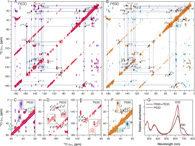
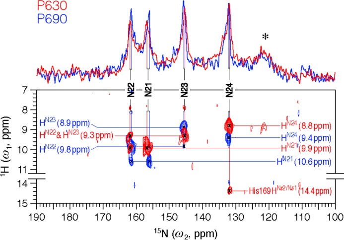
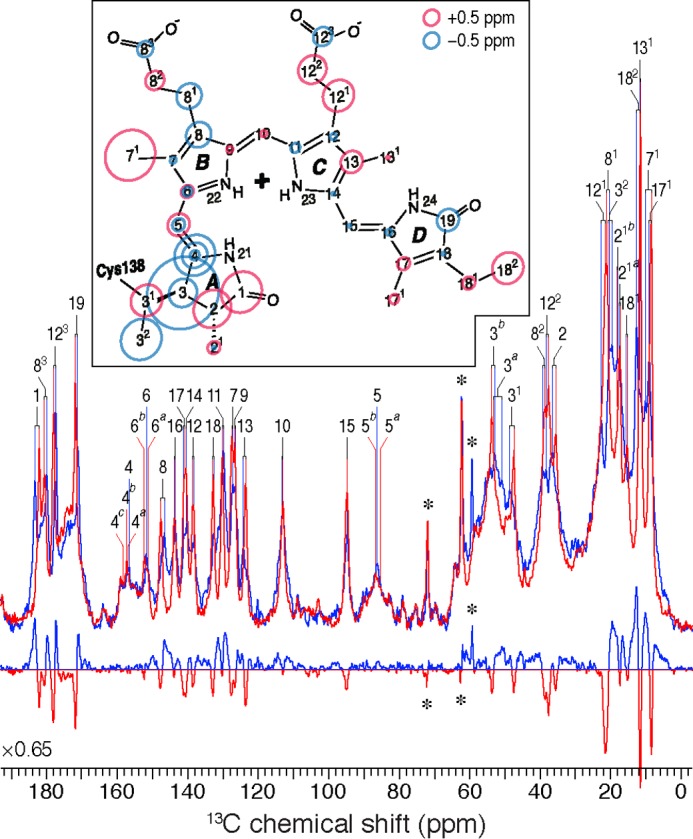
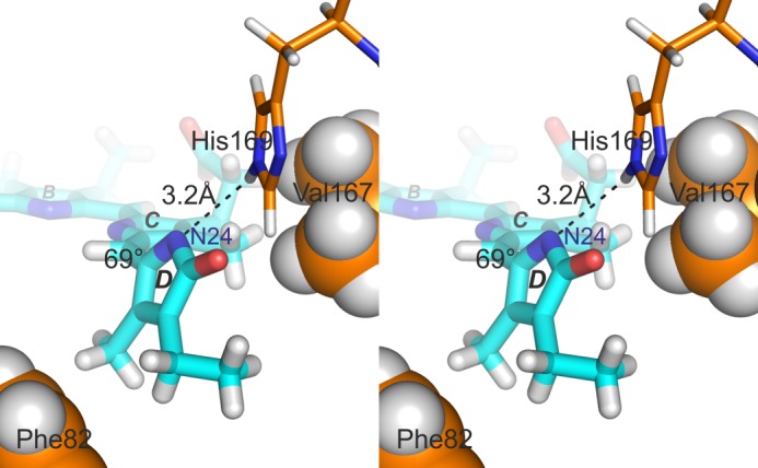
Phytochrome photoreceptors in plants and microorganisms switch photochromically between two states, controlling numerous important biological processes. Although this phototransformation is generally considered to involve rotation of ring D of the tetrapyrrole chromophore, Ulijasz et al. (Ulijasz, A. T., Cornilescu, G., Cornilescu, C. C., Zhang, J., Rivera, M., Markley, J. L., and Vierstra, R. D. (2010) Nature 463, 250-254) proposed that the A-ring rotates instead. Here, we apply magic angle spinning NMR to the two parent states following studies of the 23-kDa GAF (cGMP phosphodiesterase/adenylyl cyclase/FhlA) domain fragment of phytochrome from Synechococcus OS-B'. Major changes occur at the A-ring covalent linkage to the protein as well as at the protein residue contact of ring D. Conserved contacts associated with the A-ring nitrogen rule out an A-ring photoflip, whereas loss of contact of the D-ring nitrogen to the protein implies movement of ring D. Although none of the methine bridges showed a chemical shift change comparable with those characteristic of the D-ring photoflip in canonical phytochromes, denaturation experiments showed conclusively that the same occurs in Synechococcus OS-B' phytochrome upon photoconversion. The results are consistent with the D-ring being strongly tilted in both states and the C15=C16 double bond undergoing a Z/E isomerization upon light absorption. More subtle changes are associated with the A-ring linkage to the protein. Our findings thus disprove A-ring rotation and are discussed in relation to the position of the D-ring, photoisomerization, and photochromicity in the phytochrome family.

Keywords: Biophysics; Cyanobacteria; Molecular Biology; NMR; Photoisomerization; Photoreceptors; Phytochrome; Protein Structure; Structural Biology.


Figures

FIGURE 1.
FIGURE 1.
Two-dimensional MAS NMR 13C–13C homonuclear dipolar correlation spectra of [U-13C,15N]PCB-SyB.Cph2(GAF). Polarization transfer times of 5 and 50 ms were employed for the P630 (A, red and purple, respectively) and P690 (B, orange and cyan, respectively) states. The blue lines in two full contour plots indicate sequences of nearest-neighbor correlations (for numbering see Fig. 3, inset; for full assignments, including indirect-bonded correlations for P630 and P690 and one-dimensional spectra, see supplemental Figs. S2 and S3, respectively). The observed 13C signal splittings of a subset of carbon resonances are illustrated in C–F for P630 and P690, respectively (expansions are inset). The data were collected with an 8-ms evolution in the indirect dimension; 1434 complex t2 and 128 real t1 points with 2048 scans. A relaxation delay of 1.5 s was applied. G, UV-visible absorbance spectra of the SyB.Cph2(GAF) preparations used in this study. The spectra of P630 and of the P690/P630 photoequilibrium mixture were measured following irradiation with >700 and 525 nm light, respectively, as described.
FIGURE 2.
FIGURE 2.
Two-dimensional MAS NMR 1H–15N frequency-switched Lee-Goldburg decoupled dipolar correlation spectra of [u-13C,15N]-PCB-SyB.Cph2(GAF) as P630 (red) and P690 (blue). The regions without resonances are omitted. Two one-dimensional traces onto the 15N dimension are shown. 15N resonances (N21–N24) are indicated by vertical lines. All four NH protons (HN21-N24) are fully resolved and marked by horizontal lines. The asterisk indicates protein backbone signals in natural abundance. The spectra were acquired with 1536 complex t2 and 128 real t1 points and 2152 scans. An LG-CP contact time of 2.048 ms was applied. The relaxation delay was 1.8 s.
FIGURE 3.
FIGURE 3.
One-dimensional 13C CP/MAS NMR spectra of [U-13C,13N]PCB-SyB.Cph2(GAF) as P630 (red) and P690 (blue) and the P690–P630 difference spectrum (bottom). 13C resonances as P630 and P690 are labeled. Resonances of the natural abundance glycerol carbons are indicated by asterisks. The inset shows the changes in 13C shifts of the chromophore during photoconversion. The P630 state is taken as reference, and the size of the circles is proportional to the difference as P690–P630. Carbons showing splittings are labeled with multiple circles.
FIGURE 4.
FIGURE 4.
Stereo pair illustrating the proposed His-169···N24 hydrogen bond associated with a strongly tilted D-ring. In this model (model 5 from the corrected 2LB9 P630 ensemble, compare with Ref. 24), the hydrogen bond length is 3.2 Å and the C15=C16 dihedral angle 69°. Hydrophobic interactions with Phe-82 and Val-167 might also be important. (PyMOL image).

References

    1. Hughes J. (2010) Phytochrome three-dimensional structures and functions. Biochem. Soc. Trans. 38, 710–716 - PubMed
    1. Song C., Rohmer T., Tiersch M., Zaanen J., Hughes J., Matysik J. (2013) Solid-state NMR spectroscopy to probe photoactivation in canonical phytochromes. Photochem. Photobiol. 89, 259–273 - PubMed
    1. Rockwell N. C., Lagarias J. C. (2010) A brief history of phytochromes. ChemPhysChem 11, 1172–1180 - PMC - PubMed
    1. Wagner J. R., Brunzelle J. S., Forest K. T., Vierstra R. D. (2005) A light-sensing knot revealed by the structure of the chromophore-binding domain of phytochrome. Nature 438, 325–331 - PubMed
    1. Essen L.-O., Mailliet J., Hughes J. (2008) The structure of a complete phytochrome sensory module in the Pr ground state. Proc. Natl. Acad. Sci. U.S.A. 105, 14709–14714 - PMC - PubMed

Publication types

MeSH terms