. 2014 Jan 31;289(5):2899-907.
doi: 10.1074/jbc.M113.507491. Epub 2013 Dec 10.

The large Hsp70 Grp170 binds to unfolded protein substrates in vivo with a regulation distinct from conventional Hsp70s

Affiliations

The large Hsp70 Grp170 binds to unfolded protein substrates in vivo with a regulation distinct from conventional Hsp70s

Julia Behnke et al. J Biol Chem. .

Abstract

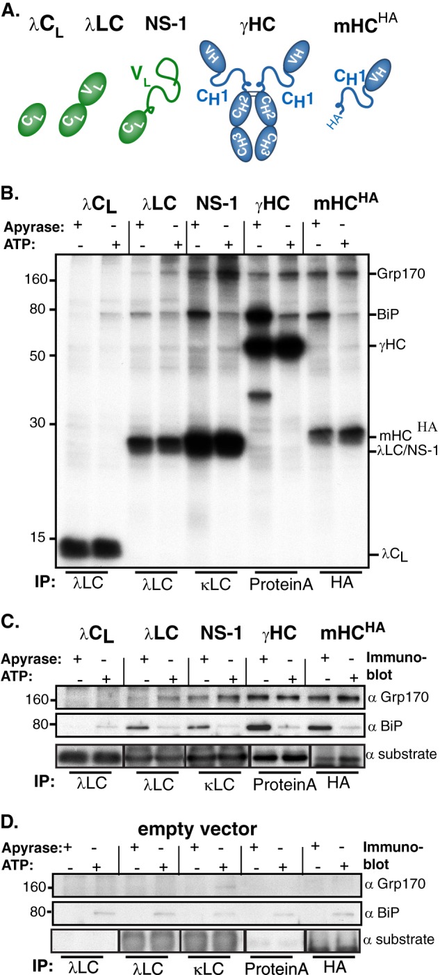
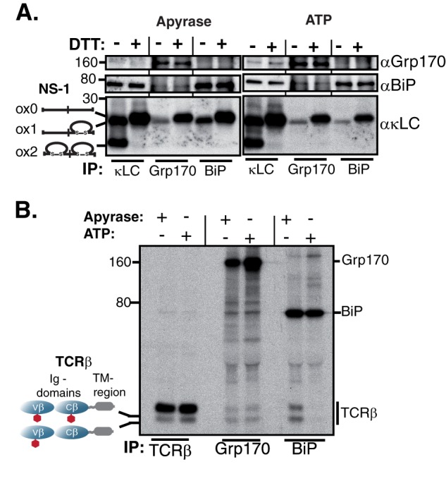
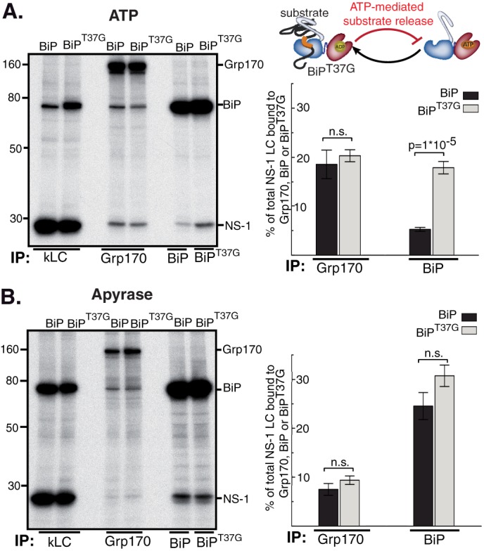
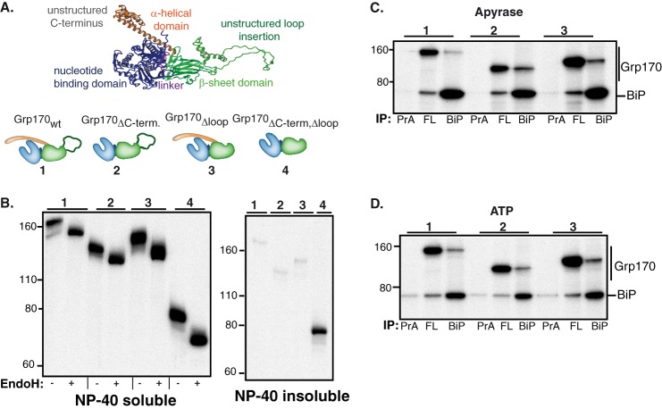
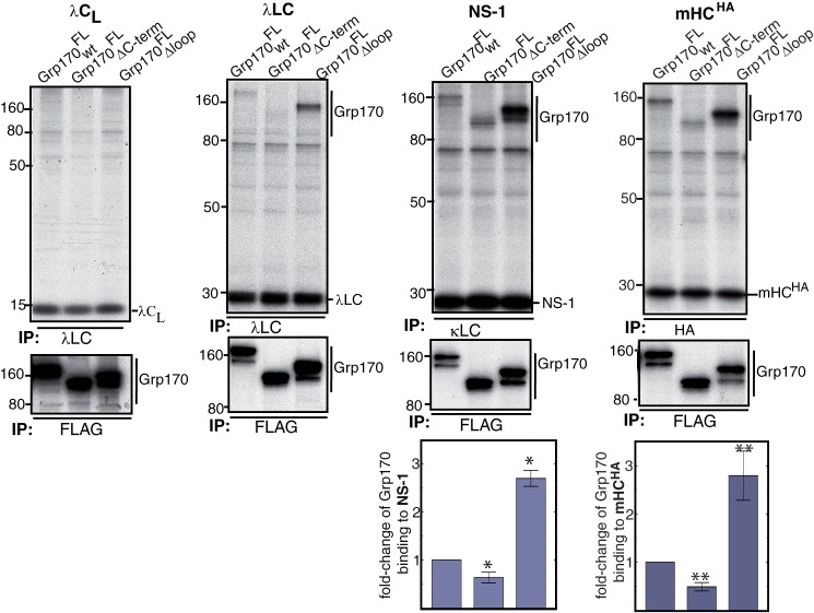
The Hsp70 superfamily is a ubiquitous chaperone class that includes conventional and large Hsp70s. BiP is the only conventional Hsp70 in the endoplasmic reticulum (ER) whose functions include: assisting protein folding, targeting misfolded proteins for degradation, and regulating the transducers of the unfolded protein response. The ER also possesses a single large Hsp70, the glucose-regulated protein of 170 kDa (Grp170). Like BiP it is an essential protein, but its cellular functions are not well understood. Here we show that Grp170 can bind directly to a variety of incompletely folded protein substrates in the ER, and as expected for a bona fide chaperone, it does not interact with folded secretory proteins. Our data demonstrate that Grp170 and BiP associate with similar molecular forms of two substrate proteins, but while BiP is released from unfolded substrates in the presence of ATP, Grp170 remains bound. In comparison to conventional Hsp70s, the large Hsp70s possess two unique structural features: an extended C-terminal α-helical domain and an unstructured loop in the putative substrate binding domain with an unknown function. We find that in the absence of the α-helical domain the interaction of Grp170 with substrates is reduced. In striking contrast, deletion of the unstructured loop results in increased binding to substrates, suggesting the presence of unique intramolecular mechanisms of control for the chaperone functions of large Hsp70s.

Keywords: ER Quality Control; Endoplasmic Reticulum (ER); Glycoprotein; Molecular Cell Biology; Molecular Chaperone; Protein Folding.


Figures

FIGURE 1.
FIGURE 1.
Grp170 directly binds to Ig substrates in vivo. A, schematic of substrates used to analyze substrate binding properties of Grp170 in vivo. Color-filled ovals represent folded Ig domains, and lines indicate unfolded regions. B, COS-1 cells were transfected with these Ig substrates along with Grp170 and BiP. Cells were pulse-labeled with [35S]cysteine/methionine for 1 h and chased for 1 h to allow maturation of the chaperones before lysing either in the presence of ATP or apyrase. Lysates were immunoprecipitated with the indicated reagents and separated by 12% SDS-PAGE followed by autoradiography. C, lysates from cells transfected as in B were immunoprecipitated with substrate specific antibodies, separated by SDS-PAGE and transferred to membranes that were blotted with either anti-Grp170, anti-BiP or anti-substrate antisera followed by species-specific secondary reagents. D, COS-1 cells were transfected and analyzed as in C, except an empty vector was transfected instead of ones encoding the substrates.
FIGURE 2.
FIGURE 2.
Grp170 and BiP bind to the same molecular species of Ig substrates. A, unlabeled P3U.1 murine myeloma cells were lysed either in the presence of apyrase or ATP, immunoprecipitated with the indicated reagents and analyzed under non-reducing conditions. As BiP is ∼10-fold more abundant than Grp170 in the cell (46), five times more lysate was used for Grp170 immunoprecipitations than for κLC or BiP. Isolated proteins were separated on 13% SDS-PAGE gels and transferred for blotting with the indicated reagents. B, COS-1 cells were transfected with vectors encoding TCRβ, Grp170, and BiP, pulse-labeled with [35S]cysteine/methionine for 0.5 h, chased for 1 h and lysed either in the presence of apyrase or ATP. Interactions between TCRβ, Grp170, and BiP were analyzed via immunoprecipitation with the indicated antisera and separation on 10% SDS gels. Four times less lysate was used for the TCRβ immunoprecipitation than for Grp170 or BiP to make both species of TCRβ visible. The mobility of the two TCRβ glycoforms is indicated.
FIGURE 3.
FIGURE 3.
Grp170 and BiP exhibit different binding kinetics to NS-1 LC. P3U.1 murine plasmacytoma cells were pulse-labeled with [35S]cysteine/methionine for 30 min followed by the indicated chase times. After lysing in the presence of apyrase, the clarified lysate was divided for immunoprecipitation with the indicated antisera. Due to different expression levels of Grp170 and BiP (46), five times more lysate was used for Grp170 immunoprecipitations. Proteins were separated by 10% SDS-PAGE and visualized by autoradiography.
FIGURE 4.
FIGURE 4.
Release from BiP is not required for substrate binding to Grp170. COS-1 cells were transfected with NS-1 LC, Grp170, and BiPWT or BiPT37G as indicated. Subsequently cells were pulse-labeled with [35S]cysteine/methionine for 0.5 h, chased for 1 h and lysed in the presence of ATP (A) or apyrase (B). The lysate was equally divided and immunoprecipitated with the indicated immune reagents. For quantitative analysis the signal of NS-1 LC bound to Grp170, BiP or BiPT37G was divided by the value for NS-1 LC obtained with κLC immunoprecipitations (n = 4 ± S.E., wild-type BiP, and n = 8 ± S.E., BiPT37G; n.s.: not significant).
FIGURE 5.
FIGURE 5.
Construction and characterization of Grp170 domain deletion mutants. A, schematic of the structural organization of Grp170 and the domain deletion mutants (blue: NBD, magenta: linker, green: β-sheet domain and unstructured loop insertion, orange: α-helical domain). The structure of human Grp170 (shown in ribbon) was modeled using Yasara Structure (www.yasara.org) based on the crystal structures of its cytosolic yeast orthologue Sse1p (13, 14, 40) and used to design FLAG-tagged Grp170 whole domain deletion mutants, which are numbered. B, COS-1 cells were transfected with empty pSVL vector, BiP, and the indicated Grp170 constructs. After a 1 h pulse-label with [35S]cysteine/methionine and a 1 h chase, cells were lysed in the presence of ATP. After centrifugation, samples were divided into Nonidet P-40 soluble and Nonidet P-40 insoluble fraction and immunoprecipitated with antiserum against the FLAG-tag. The eluted protein from cell lysates was divided and left undigested or treated with Endo H. Samples were analyzed by 10% SDS-PAGE, followed by autoradiography. The numbers above each group correspond to the deletion mutants shown in A. C and D, COS-1 cells were transfected with BiP and the indicated Grp170 constructs. After labeling with [35S]cysteine/methionine for 5 h and a chase period of 16 h, cells were lysed in the presence of apyrase (C) or ATP (D). Cell lysates were divided equally and immunoprecipitated with an antiserum against either the FLAG-tag (FL), BiP, or protein A (PrA) only. The proteins were separated on 10% SDS-PAGE, followed by autoradiography. Deletion mutants are indicated by the number above each group.
FIGURE 6.
FIGURE 6.
Grp170's unstructured loop and C-terminal α-helical domain modulate substrate binding. COS-1 cells were transfected with FLAG-tagged Grp170 constructs, BiP and the indicated Ig proteins. Following a 1 h pulse-label with [35S]cysteine/methionine and 1 h chase, cells were lysed in the presence of ATP, and the cell lysate was equally divided for immunoprecipitation with substrate-specific antisera or FLAG-tag as indicated. Proteins were separated on 10% SDS-gels, followed by autoradiography. The signals of Grp170 constructs bound to NS-1 and mHCHA were quantified and corrected for the Grp170 and substrate expression levels. The value obtained for Grp170wtFL was set to 1 (NS-1 LC: n = 7 ± S.E., *, p ≤ 0.007; mHCHA: n = 3 ± S.E., **, p ≤ 0.02).

References

    1. Braakman I., Bulleid N. J. (2011) Protein folding and modification in the mammalian endoplasmic reticulum. Annu. Rev. Biochem. 80, 71–99 - PubMed
    1. Hartl F. U., Bracher A., Hayer-Hartl M. (2011) Molecular chaperones in protein folding and proteostasis. Nature 475, 324–332 - PubMed
    1. Hebert D. N., Molinari M. (2012) Flagging and docking: dual roles for N-glycans in protein quality control and cellular proteostasis. Trends Biochem. Sci 37, 404–410 - PMC - PubMed
    1. Otero J. H., Lizák B., Hendershot L. M. (2010) Life and death of a BiP substrate. Semin Cell Dev. Biol. 21, 472–478 - PMC - PubMed
    1. Kampinga H. H., Craig E. A. (2010) The HSP70 chaperone machinery: J proteins as drivers of functional specificity. Nat. Rev. Mol. Cell Biol. 11, 579–592 - PMC - PubMed

Publication types

MeSH terms

LinkOut - more resources