. 2014 Jan 8;136(1):137-46.
doi: 10.1021/ja411366w. Epub 2013 Dec 20.

Human cellular retinaldehyde-binding protein has secondary thermal 9-cis-retinal isomerase activity

Affiliations

Human cellular retinaldehyde-binding protein has secondary thermal 9-cis-retinal isomerase activity

Christin S Bolze et al. J Am Chem Soc. .

Abstract

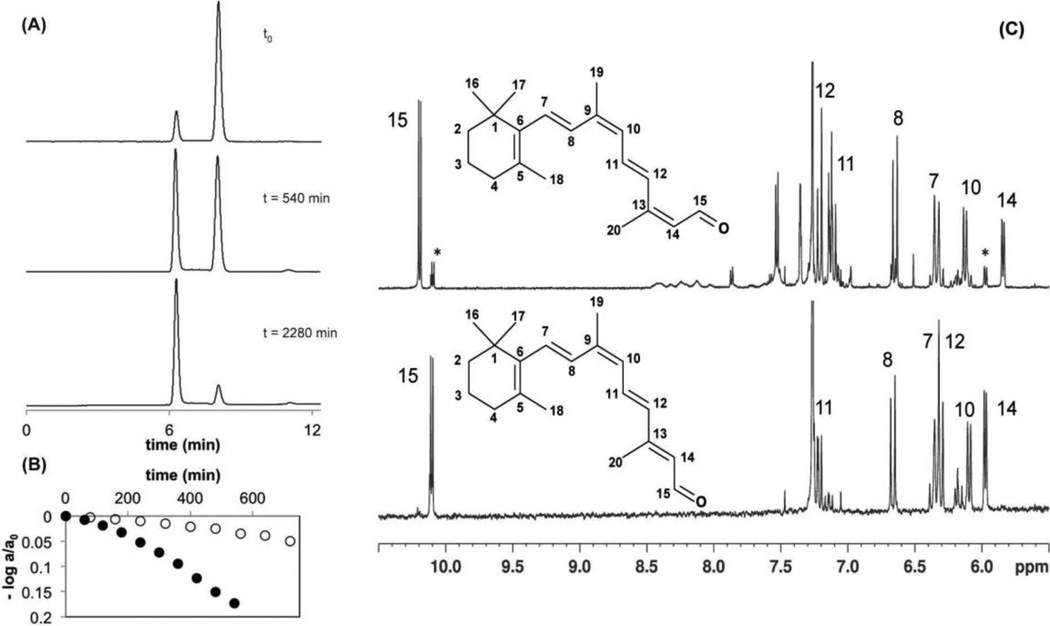
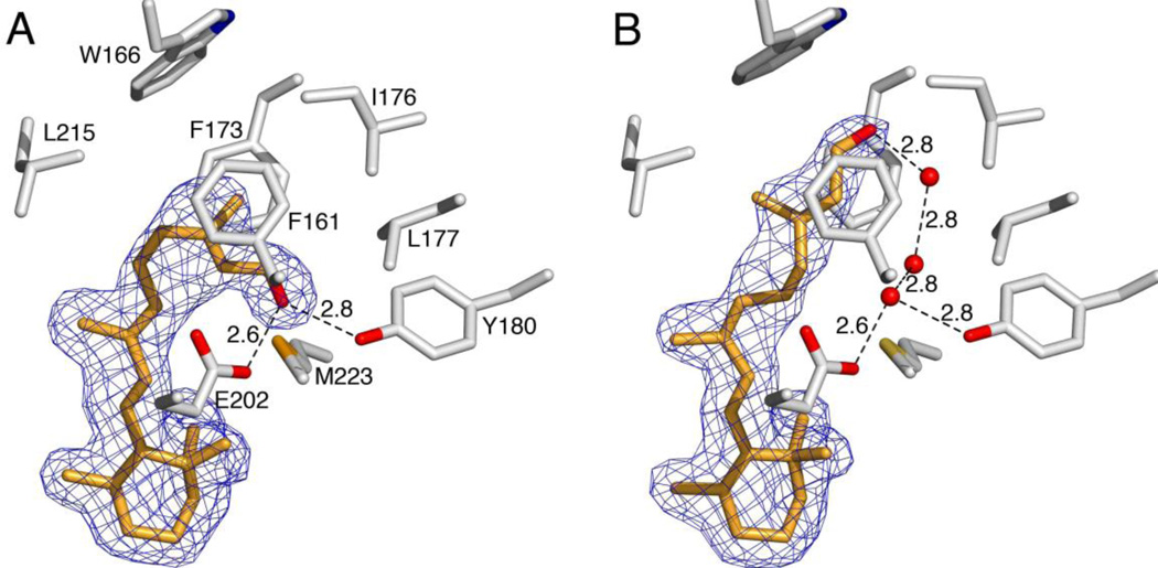
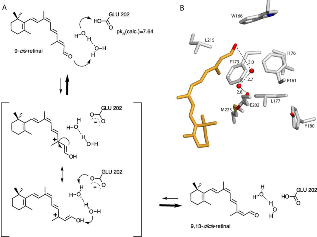
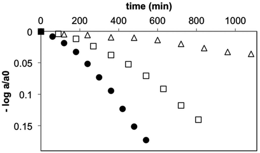
Cellular retinaldehyde-binding protein (CRALBP) chaperones 11-cis-retinal to convert opsin receptor molecules into photosensitive retinoid pigments of the eye. We report a thermal secondary isomerase activity of CRALBP when bound to 9-cis-retinal. UV/vis and (1)H NMR spectroscopy were used to characterize the product as 9,13-dicis-retinal. The X-ray structure of the CRALBP mutant R234W:9-cis-retinal complex at 1.9 Å resolution revealed a niche in the binding pocket for 9-cis-aldehyde different from that reported for 11-cis-retinal. Combined computational, kinetic, and structural data lead us to propose an isomerization mechanism catalyzed by a network of buried waters. Our findings highlight a specific role of water molecules in both CRALBP-assisted specificity toward 9-cis-retinal and its thermal isomerase activity yielding 9,13-dicis-retinal. Kinetic data from two point mutants of CRALBP support an essential role of Glu202 as the initial proton donor in this isomerization reaction.


Figures

Figure 1
Figure 1. Characterization of a CRALBP:9-cis-retinal reaction product
A) HPLC traces of retinoid extracts of CRALBP:9-cis-retinal freshly prepared (top), after 9 h incubation at 37°C (middle) and after 38 h incubation at 37°C (bottom). B) Kinetics of CRALBP:9-cis-retinal isomerization in H2O buffer (filled circles) and in D2O buffer (open circles) quantified by HPLC and plotted versus time. Rate constants (k) were determined from the fit of the data to the following equation: log (A380(t)/A380(t0)) = −kt/ln(10), where ΔA(t) = A380(t = 0) −A380(t). C) 500 MHz 1H-NMR spectra of 9,13-dicis-retinal (top) and 9-cis-retinal (bottom) produced by incubation of 9-cis-retinal in CRALBP. Both retinoids were extracted from the complex and dissolved in CDCl3. Allocations of the six vinyl protons 7, 8, 10, 11, 12, and 14, as well as of the aldehyde proton 15 are indicated in the spectra, according to the numbering given in the structures. A second set of resonances visible for protons 14 and 15 and marked with asterisks, indicates traces of residual 9-cis-retinal.
Figure 2
Figure 2. Close-up views of the R234W ligand-binding pockets
A) R234W bound to 11-cis-retinal. B) R234W bound to 9-cis-retinal. Side chains (gray) that form van der Waals contacts with the cis-retinoid tails (orange) or participate in hydrogen bonding within the ligand-binding pockets are shown as sticks. The 2Fo–Fc electron densities for the ligands are shown as blue mesh at 1 σ. Dashed black lines indicate hydrogen bonds; distances are given in A.
Figure 3
Figure 3. Superposition of different computational models of 9-cis-retinal in R234W
The retinal model fitting the experimental X-ray density is shown in cyan balls-and-sticks. The QM/MM optimized geometry obtained for the neutral closed-shell molecule is drawn in silver licorice; the carbocation with H+ attached at C12 in orange licorice; and the optimized structure of the radical obtained by H capture at C12 in blue licorice.
Figure 4
Figure 4. Mechanism of the isomerization reaction
A) Proposed mechanism for the thermal isomerization of 9-cis-retinal to 9,13-dicis-retinal in CRALBP. The reaction proceeds via direct protonation of the polyene aldehyde, isomerization of the charge separated transition state and final deprotonation of the enol. B) Close-up view of the MD structural model of the dark state binding pocket of CRALBP:9-cis-retinal. Side chains and 9-cis-retinal are shown as gray and orange sticks, respectively. Dashed black lines indicate hydrogen bonds. Distances are given in A.
Figure 5
Figure 5. Comparison of the reaction kinetics of CRALBP, E202Q and Y180F
Kinetics of CRALBP:9-cis-retinal isomerization (filled circles), E202Q (open triangles) and Y180F (open squares) quantified by HPLC and plotted versus time. Rate constants (k) were determined from fitting the data to the following equation: log (A380(t)/A380(t0)) = −kt/ln(10), where ΔA(t) = A380(t = 0) − A380(t). k = 1.1×10⁻⁵s−1 (CRALBP:9-cis-retinal); k = 0.10×10⁻⁵ s−1, (E202Q), and k = 0.57×10⁻⁵ s−1 (Y180F).

References

    1. Palczewski K. J. Biol. Chem. 2012;287:1612–1619. - PMC - PubMed
    1. Palczewski K. Annu. Rev. Biochem. 2006;75:743–767. - PMC - PubMed
    1. Travis GH, Golczak M, Moise AR, Palczewski K. Annu. Rev. Pharmacol. Toxicol. 2007;47:469–512. - PMC - PubMed
    1. Fleisch VC, Neuhauss SC. Prog. Ret. Eye Res. 2010;29:476–486. - PubMed
    1. Moiseyev G, Chen Y, Takahashi Y, Wu BX, Ma JX. Proc. Natl. Acad. Sci. U. S. A. 2005;102:12413–12418. - PMC - PubMed

Publication types