. 2014 Mar;164(6):811-21.
doi: 10.1111/bjh.12708. Epub 2013 Dec 13.

Immunomodulatory agents lenalidomide and pomalidomide co-stimulate T cells by inducing degradation of T cell repressors Ikaros and Aiolos via modulation of the E3 ubiquitin ligase complex CRL4(CRBN.)

Affiliations

Immunomodulatory agents lenalidomide and pomalidomide co-stimulate T cells by inducing degradation of T cell repressors Ikaros and Aiolos via modulation of the E3 ubiquitin ligase complex CRL4(CRBN.)

Anita K Gandhi et al. Br J Haematol. 2014 Mar.

Abstract

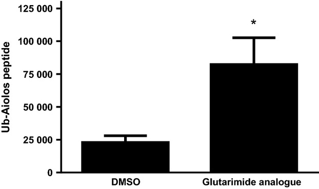
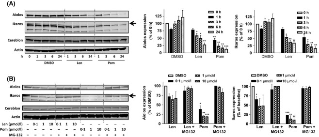
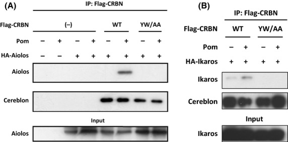
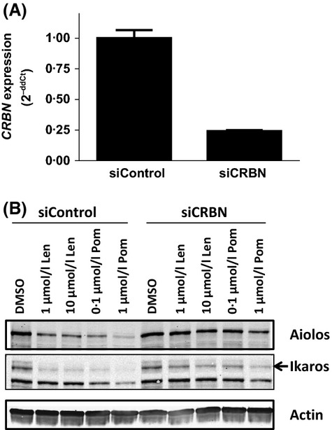
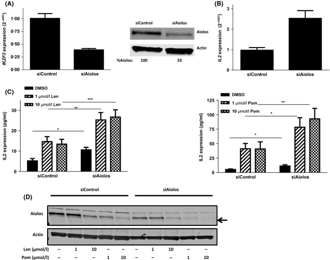
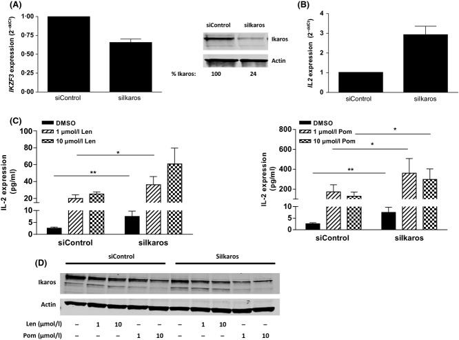
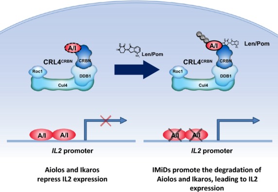
Cereblon (CRBN), the molecular target of lenalidomide and pomalidomide, is a substrate receptor of the cullin ring E3 ubiquitin ligase complex, CRL4(CRBN) . T cell co-stimulation by lenalidomide or pomalidomide is cereblon dependent: however, the CRL4(CRBN) substrates responsible for T cell co-stimulation have yet to be identified. Here we demonstrate that interaction of the transcription factors Ikaros (IKZF1, encoded by the IKZF1 gene) and Aiolos (IKZF3, encoded by the IKZF3 gene) with CRL4(CRBN) is induced by lenalidomide or pomalidomide. Each agent promotes Aiolos and Ikaros binding to CRL4(CRBN) with enhanced ubiquitination leading to cereblon-dependent proteosomal degradation in T lymphocytes. We confirm that Aiolos and Ikaros are transcriptional repressors of interleukin-2 expression. The findings link lenalidomide- or pomalidomide-induced degradation of these transcriptional suppressors to well documented T cell activation. Importantly, Aiolos could serve as a proximal pharmacodynamic marker for lenalidomide and pomalidomide, as healthy human subjects administered lenalidomide demonstrated Aiolos degradation in their peripheral T cells. In conclusion, we present a molecular model in which drug binding to cereblon results in the interaction of Ikaros and Aiolos to CRL4(CRBN) , leading to their ubiquitination, subsequent proteasomal degradation and T cell activation.

Keywords: Aiolos; Cereblon; Ikaros; lenalidomide; pomalidomide.


Figures

Figure 1
Figure 1
A ubiquitinated Aiolos peptide is enriched in T cells treated with a Cereblon-binding glutarimide analogue. T cells from multiple donors were treated with DMSO or drug for 6 h followed by enrichment for ubiquitinated peptides. Peptides were analysed by LC-MS/MS and data shown are mean Ub-Aiolos peptide peak height ± SD; *< 0·05 versus DMSO using paired t-test. DMSO, dimethyl sulfoxide; Ub, ubiquitinated; SD, standard deviation.
Figure 2
Figure 2
Lenalidomide and pomalidomide induce the degradation of Aiolos and Ikaros in T cells in a time-dependent manner and concentration-dependent manner via proteosomal degradation. (A) T cells were treated with 10 μmol/l lenalidomide or 1 μmol/l pomalidomide for 1, 3, 6 and 24 h. Lysates were separated by SDS-PAGE and immunoblotted for Aiolos, Ikaros, Cereblon and Actin expression; left representative image, middle densitometry analysis of Aiolos, right densitometry analysis of Ikaros expression normalized to Actin; (B) T cells were pre-treated with MG-132 for 30 min then treated with lenalidomide or pomalidomide for 6 h at the indicated concentrations. Lysates were separated by SDS-PAGE and immunoblotted for Aiolos, Ikaros, Cereblon and Actin expression; left representative image, middle densitometry analysis of Aiolos, right densitometry analysis of Ikaros expression normalized to Actin; data shown is mean ± SEM of four experiments; *< 0·05, **< 0·01 and ***< 0·001 using one-way anova. Len, lenalidomide; Pom, pomalidomide; SDS-PAGE, sodium dodecyl sulphate polyacrylamide gel electrophoresis; SEM, standard error of the mean; anova, analysis of variance.
Figure 3
Figure 3
Cereblon binds Aiolos and Ikaros and requires the thalidomide binding domain on Cereblon. (A) HEK-293T cells were transfected with Flag-WT Cereblon or Flag-YW/AA Cereblon with or without HA-Aiolos for 48 h with pomalidomide added to cells for the last hour. Lysates were made and immunoprecipitation with Flag antibody followed by SDS-PAGE separation and immunoblotting with anti-Aiolos or anti–Cereblon was performed. (B) HEK-293T cells were transfected with Flag-WT Cereblon or Flag-YW/AA Cereblon with or without HA-Ikaros for 48 h with pomalidomide added to cells for the last hour. Lysates were made and immunoprecipitation with Flag antibody followed by SDS-PAGE separation and immunoblotting with anti–Cereblon or anti-Ikaros was performed. IP, immunoprecipitation; CRBN, Cereblon; WT, wildtype; SDS-PAGE, sodium dodecyl sulphate polyacrylamide gel electrophoresis.
Figure 4
Figure 4
Lenalidomide and pomalidomide stimulate Aiolos degradation via ubiquitination and protein turnover. (A) Jurkat cells were transfected with control empty vector, wildtype IKZF3 or mutant IKZF3 for 6 h then treated with either DMSO, 1 or 10 μmol/l lenalidomide or pomalidomide for 24 h. Cell lysates were separated by SDS-PAGE and immunoblotted for Aiolos and Actin. (B) U266 cells were pulsed with 10 μg/ml cycloheximide and treated with either DMSO, 10 μmol/l lenalidomide or 1 μmol/l pomalidomide for 1·5, 3 or 6 h. Cell lysates were separated by SDS-PAGE and immunoblotted for Aiolos, Ikaros and Actin. Densitometry analysis of Aiolos and Ikaros expression normalized to Actin; data shown is mean ± SEM of three experiments. Len, lenalidomide; Pom, pomalidomide; CHX, cycloheximide; Mt, mutant; WT, wildtype; DMSO, dimethyl sulfoxide; SDS-PAGE, sodium dodecyl sulphate polyacrylamide gel electrophoresis.
Figure 5
Figure 5
Aiolos and Ikaros degradation by lenalidomide and pomalidomide is Cereblon-dependent. (A) Primary human CD3+ T cells were transfected with control siRNA or CRBN siRNA for 48 h and CRBN knockdown efficiency was measured 24 h post-transfection by qRT-PCR. (B) CRBN siRNA knockdown T cells were treated with lenalidomide or pomalidomide at the indicated concentrations for 24 h. Cell lysates were separated by SDS-PAGE and immunoblotted for Aiolos, Ikaros and Actin expression. Len, lenalidomide; Pom, pomalidomide; CRBN, Cereblon gene; siCRBN, CRBN siRNA; SDS-PAGE, sodium dodecyl sulphate polyacrylamide gel electrophoresis.
Figure 6
Figure 6
Aiolos is a negative regulator of IL2 in T cells and silencing Aiolos mimics lenalidomide or pomalidomide treatment. (A) Primary T cells were transfected with control siRNA or Aiolos siRNA for 24 h then assayed for IKZF3 gene expression or lysates were separated by SDS-PAGE and immunoblotted for Aiolos and Actin expression. Values represent Aiolos densitometry values normalized to Actin. (B) T cells transfected with control siRNA or Aiolos siRNA for 24 h were assayed for IL2 gene expression. (C) 24 h post-siRNA transfection, T cells were treated with drug for 48 h (left lenalidomide, right pomalidomide), harvested and IL2 protein expression was measured by ELISA. Data shown are mean of three donors tested in triplicate. (D) Aiolos and Actin expression was measured after drug treatment by immunoblot. *< 0·05. **< 0·01 and ***< 0·001 using paired t-test. Len, lenalidomide; Pom, pomalidomide; siAiolos, Aiolos siRNA; DMSO, dimethyl sulfoxide; SDS-PAGE, sodium dodecyl sulphate polyacrylamide gel electrophoresis; ELISA, enzyme-linked immunosorbent assay.
Figure 7
Figure 7
Ikaros is a negative regulator of IL2 in T cells and silencing Ikaros mimics lenalidomide or pomalidomide treatment. (A) Primary T cells were transfected with Control siRNA or Ikaros siRNA for 48 h then assayed for IKZF1 gene expression or lysates were separated by SDS-PAGE and immuno blotted for Ikaros expression. Values represent Ikaros densitometry values normalized to Actin. (B) T cells transfected with control or Ikaros siRNA for 24 h were assayed for IL2 gene expression (n = 4). (C) 24 h post-siRNA transfection, T cells were drug treated for 48 h (left, lenalidomide; right, pomalidomide), harvested and IL2 expression was measured by ELISA. Data shown are mean of three donors tested in triplicate. (D) Ikaros and Actin expression was measured by immunoblot after drug treatment. *< 0·05. and **< 0·01 using paired t-test. Len, lenalidomide; Pom, pomalidomide; siIkaros, Ikaros siRNA; DMSO, dimethyl sulfoxide; SDS-PAGE, sodium dodecyl sulphate polyacrylamide gel electrophoresis; ELISA, enzyme-linked immunosorbent assay.
Figure 8
Figure 8
Dose-dependent Aiolos inhibition in peripheral T cells from healthy volunteers administered with lenalidomide. Healthy volunteers were administered either placebo, 10 or 50 mg of lenalidomide and blood was drawn 6 h post-dose. Aiolos levels were measured in CD3+ T cells by flow cytometry (n = 5–8 for each treatment arm). MEFL, mean equivalent fluorochrome labelling; SEM, standard error of the mean.
Figure 9
Figure 9
Model of lenalidomide and pomalidomide Co-stimulation of T cells via Degradation of Aiolos and Ikaros. In the absence of lenalidomide and pomalidomide, Aiolos and Ikaros are expressed and function as repressors of the IL2 promoter. Aiolos and Ikaros may or may not be normal substrates of Cereblon as represented by the dashed line, however the presence of lenalidomide and pomalidomide increases the ability of CRL4CRBN to interact with Aiolos and Ikaros, leading to their ubiquitination and degradation resulting in de-repression of the IL2 promoter and increased expression of IL2. CRL4, cullin ring 4 ligase; CRBN, Cereblon; A/I, Aiolos/Ikaros; Cul4, cullin 4; DDB1, DNA damage binding protein-1; IMiD, immunomodulatory drug; Len, lenalidomide; Pom, pomalidomide; Roc1, regulator of cullins 1; Ub, ubiquitin.

References

    1. Bandyopadhyay S, Duré M, Paroder M, Soto-Nieves N, Puga I. Macián F. Interleukin 2 gene transcription is regulated by Ikaros-induced changes in histone acetylation in anergic T cells. Blood. 2007;109:2878–2886. - PMC - PubMed
    1. Billot K, Parizot C, Arrouss I, Mazier D, Debre P, Rogner UC. Rebollo A. Differential Aiolos expression in human hematopoietic subpopulations. Leukemia Research. 2010;34:289–293. - PubMed
    1. Cai Q, Dierich A, Oulad-Abdelghani M, Chan S. Kastner P. Helios deficiency has minimal impact on T cell development and function. Journal of Immunology. 2009;183:2303–2311. - PubMed
    1. Chen N, Lau H, Kong L, Kumar G, Zeldis JB, Knight R. Laskin OL. Pharmacokinetics of lenalidomide in subjects with various degrees of renal impairment and in subjects on hemodialysis. Journal of Clinical Pharmacology. 2007;47:1466–1475. - PubMed
    1. Corral LG, Haslett PA, Muller GW, Chen R, Wong LM, Ocampo CJ, Patterson RT, Stirling DI. Kaplan G. Differential cytokine modulation and T cell activation by two distinct classes of thalidomide analogues that are potent inhibitors of TNF-α. Journal of Immunology. 1999;163:380–386. - PubMed

MeSH terms