. 2014 Apr;171(7):1655-67.
doi: 10.1111/bph.12552.

Mechanism of cytotoxic action of crambescidin-816 on human liver-derived tumour cells

Affiliations

Mechanism of cytotoxic action of crambescidin-816 on human liver-derived tumour cells

J A Rubiolo et al. Br J Pharmacol. 2014 Apr.

Abstract

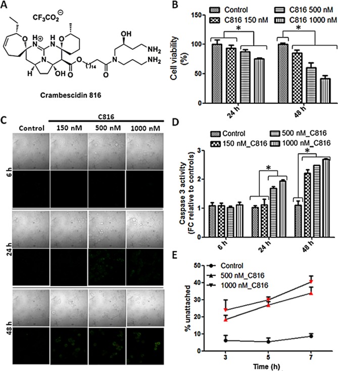
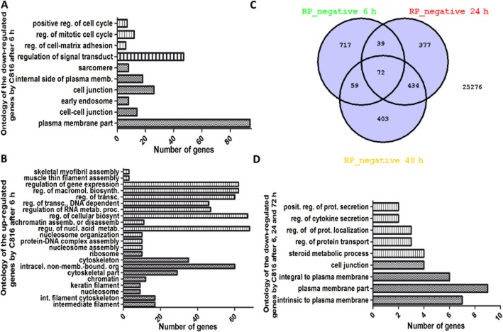
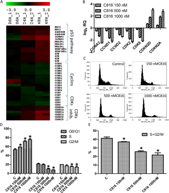
Background and purpose: Marine sponges have evolved the capacity to produce a series of very efficient chemicals to combat viruses, bacteria, and eukaryotic organisms. It has been demonstrated that several of these compounds have anti-neoplastic activity. The highly toxic sponge Crambe crambe has been the source of several molecules named crambescidins. Of these, crambescidin-816 has been shown to be cytotoxic for colon carcinoma cells. To further investigate the potential anti-carcinogenic effect of crambescidin-816, we analysed its effect on the transcription of HepG2 cells by microarray analysis followed by experiments guided by the results obtained.

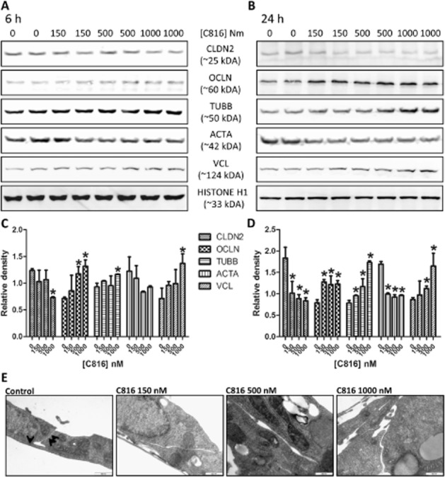
Experimental approach: After cytotoxicity determination, a transcriptomic analysis was performed to test the effect of crambescidin-816 on the liver-derived tumour cell HepG2. Based on the results obtained, we analysed the effect of crambescidin-816 on cell-cell adhesion, cell-matrix adhesion, and cell migration by Western blot, confocal microscopy, flow cytometry and transmission electron microscopy. Cytotoxicity and cell migration were also studied in a variety of other cell lines derived from human tumours.

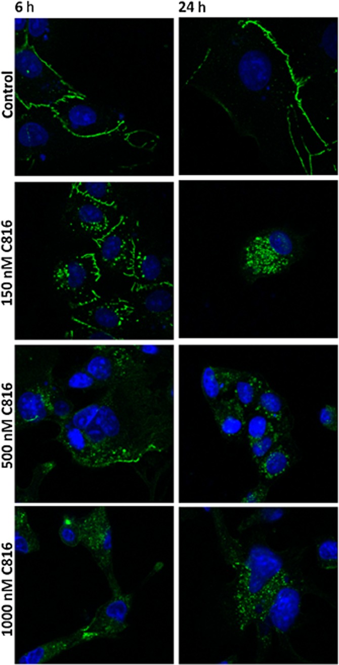
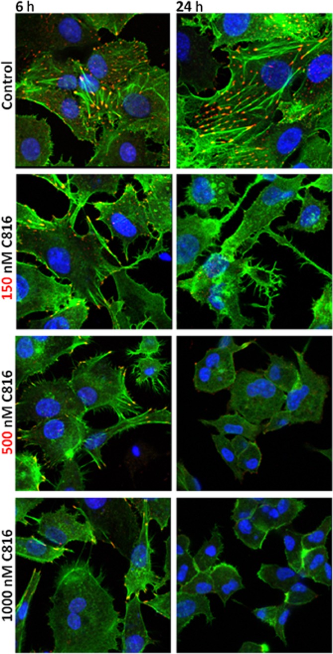
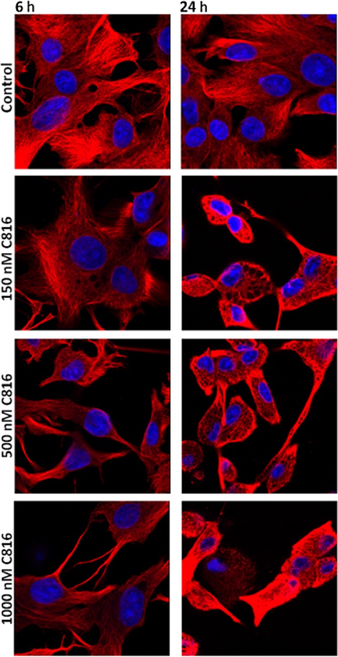
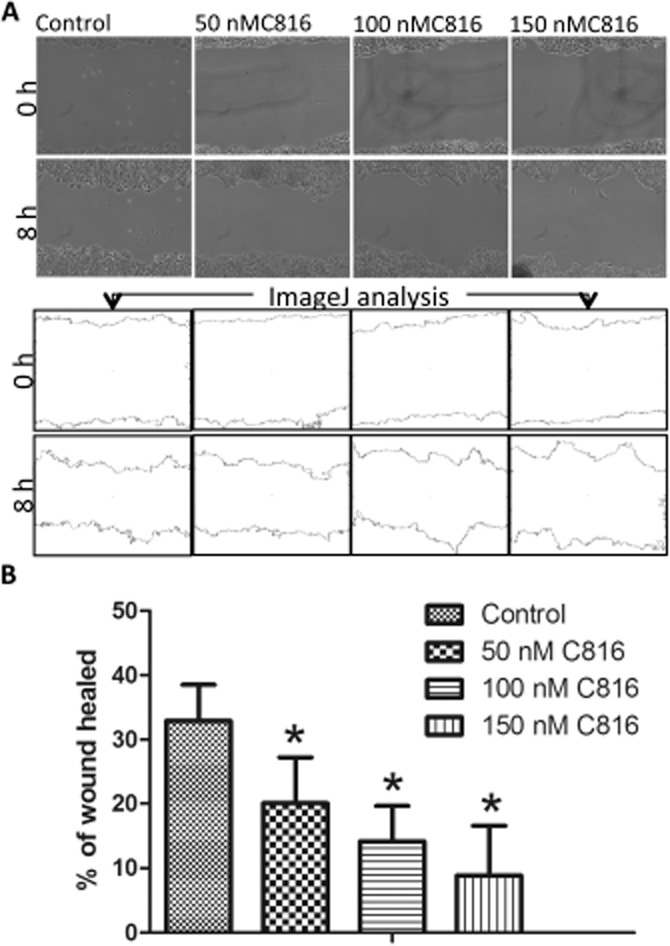
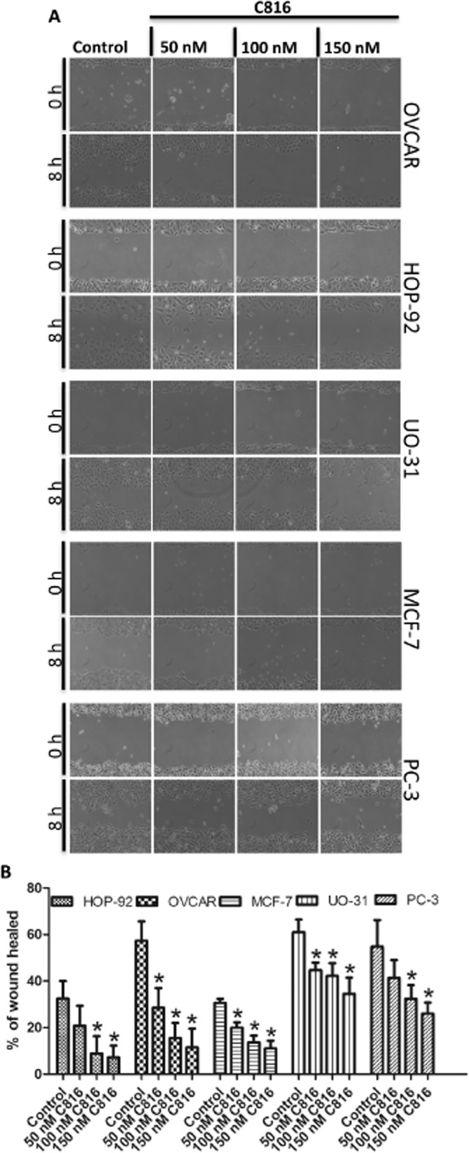
Key results: Crambescidin-816 had a cytotoxic effect on all the cell lines studied. It inhibited cell-cell adhesion, interfered with the formation of tight junctions, and cell-matrix adhesion, negatively affecting focal adhesions. It also altered the cytoskeleton dynamics. As a consequence of all these effects on cells crambescidin-816 inhibited cell migration.

Conclusions and implications: The results indicate that crambescidin-816 is active against tumour cells and implicate a new mechanism for the anti-tumour effect of this compound.

Keywords: crambescidin-816; transcriptomic analysis; tumour cell adhesion; tumour cell migration; tumour cell viability.


Figures

Figure 1
Figure 1
(A) Structure of C816. (B) Viability of HepG2 cells after C816 treatment for 24 and 48 h, determined by the MTT method. P < 0.01, n = 8. (C) Annexin V and PI staining of HepG2 cells after C816 treatment for 6, 24 and 48 h. Representative photos of the merged Annexin V and PI fluorescence together with bright field for each treatment are shown. (D) Caspase-3 activity in HepG2 cells treated with C816 for 6, 24 and 48 h. *Significant differences with respect to controls, P < 0.01, n = 3. FC: fold change. (E) Quantification of unattached cells in bright field images of control and C816-treated HepG2 cells. Seven fields from each treatment were analysed. Red symbols: significant differences with respect to controls, P < 0.01, n = 7.
Figure 2
Figure 2
Gene ontology of the down-regulated (A) and up-regulated (B) biological processes and cell components in HepG2 cells after 6 h of C816 treatment. Hatched bars: biological processes level 5. Stippled bars: cell components level 5. (C) Venn diagram for the down-regulated mRNAs in HepG2 cells after C816 treatment. (D) Gene ontology of the down-regulated genes shared by the three time points analysed. Hatched bars: biological processes level 5. Stippled bars: cell components level 5.
Figure 3
Figure 3
(A) Heat map of the DE expressed mRNAs (in log2) coding for proteins involved in cell cycle regulation in 150 nM C816-treated cells with respect to controls, determined by microarray data analysis. Green: mRNA down-regulation in treated cells with respect to controls. Red: mRNA up-regulation in treated cells with respect to controls. (B) qPCR validation of the microarray (48 h) data for cyclins A and D, CDKs 2 and 6, and CDK inhibitors 2D and 2A, and determination of the effect of higher C816 concentrations. (C) Representative histograms of the cell cycle obtained after flow cytometry analysis of C816-treated HepG2 cells for 24 h. (D) Quantification of the cell populations in each phase of the cell cycle in control and C816-treated (24 h) HepG2 cells (P < 0.01, n = 3). (E) Quantification of the overall effect on S and G2/M phases produced by C816 after 24 h on HepG2 cells (P < 0.01, n = 3).
Figure 4
Figure 4
Detection of CLDN2, OCLN, ACTA, TUBB, VNC and histone H1 by Western blot. The experiment was performed using soluble protein obtained from HepG2 cells after treatment with 150, 500 and 1000 nM C816 for 6 h (A) and 24 h (B). Quantification of the variations in the proteins assayed by Western blot between controls and treated cells after 6 h (C) and 24 h (D). (E) TEM of HepG2 cells treated with 150, 500 and 1000 nM C816 for 24 h showing the increased separation between cell membranes in treated cells. Arrows: tight junctions.
Figure 5
Figure 5
OCLN detection (green) by confocal microscopy in control and C816-treated HepG2 cells. Representative photos of treatments for 6 and 24 h are shown. In both cases, nuclei were counterstained with Hoechst 33258.
Figure 6
Figure 6
Actin (green) and vinculin (red) detection by confocal microscopy in control and C816-treated HepG2 cells. Representative photos of treatments for 6 and 24 h are shown. Colocalization of actin and vinculin is shown in yellow. In both cases, nuclei were counterstained with Hoechst 33258.
Figure 7
Figure 7
β-Tubulin detection by confocal microscopy in control and C816-treated HepG2 cells. Representative photos of treatments for 6 and 24 h are shown. In both cases, nuclei were counterstained with Hoechst 33258.
Figure 8
Figure 8
(A) Representative photos and ImageJ analysis to determine the percentage of wound healed in HepG2 cells after treatment with 50, 100 and 150 nM C816 for 8 h. (B) Quantification of wound healing after 8 h in HepG2 cells treated with 50, 100 and 150 nM C816 (P < 0.01, n = 20).
Figure 9
Figure 9
(A) Representative photos obtained in the wound healing assays after treatment of HOP-92, MCF-7, OVCAR, UO-31 and PC-3 cells for 8 h with C816. (B) Quantification of wound healing after 8 h of exposure to 50, 100 and 150 nM C816 (P < 0.01, n = 20).

References

    1. Aoki S, Kong D, Matsui K, Kobayashi M. Erythroid differentiation in K562 chronic myelogenous cells induced by crambescidin 800, a pentacyclic guanidine alkaloid. Anticancer Res. 2004;24:2325–2330. - PubMed
    1. Aron ZD, Pietraszkiewicz H, Overman LE, Valeriote F, Cuevas C. Synthesis and anticancer activity of side chain analogs of the crambescidin alkaloids. Bioorg Med Chem Lett. 2004;14:3445–3449. - PubMed
    1. Bates S, Bonetta L, MacAllan D, Parry D, Holder A, Dickson C, et al. CDK6 (PLSTIRE) and CDK4 (PSK-J3) are a distinct subset of the cyclin-dependent kinases that associate with cyclin D1. Oncogene. 1994;9:71–79. - PubMed
    1. Belarbi el H, Contreras Gomez A, Chisti Y, Garcia Camacho F, Molina Grima E. Producing drugs from marine sponges. Biotechnol Adv. 2003;21:585–598. - PubMed
    1. Berlinck RG, Braekman JC, Daloze D, Bruno I, Riccio R, Ferri S, et al. Polycyclic guanidine alkaloids from the marine sponge Crambe crambe and Ca++ channel blocker activity of crambescidin 816. J Nat Prod. 1993;56:1007–1015. - PubMed

Publication types

MeSH terms