. 2014 Feb;20(3-4):774-84.
doi: 10.1089/ten.TEA.2013.0122. Epub 2014 Jan 17.

Epithelial-differentiated adipose-derived stem cells seeded bladder acellular matrix grafts for urethral reconstruction: an animal model

Affiliations

Epithelial-differentiated adipose-derived stem cells seeded bladder acellular matrix grafts for urethral reconstruction: an animal model

Hongbin Li et al. Tissue Eng Part A. 2014 Feb.

Abstract

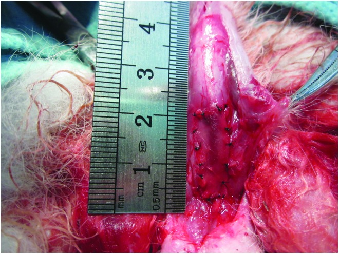
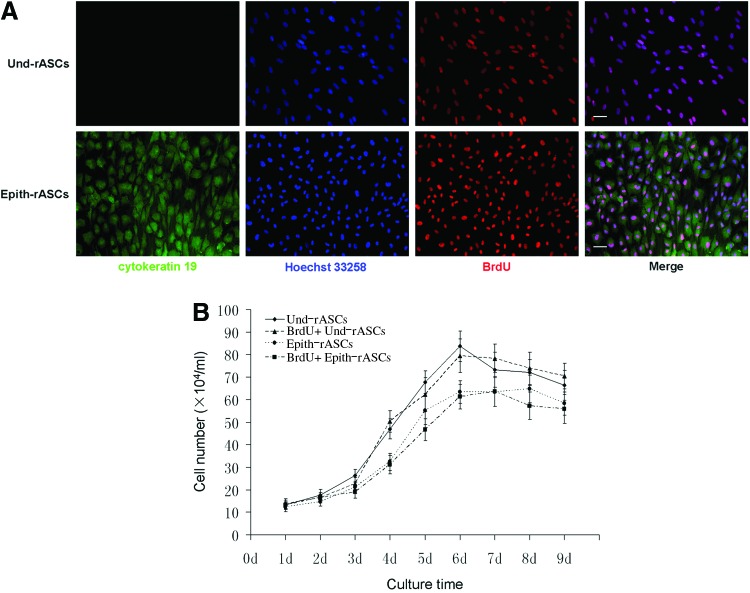
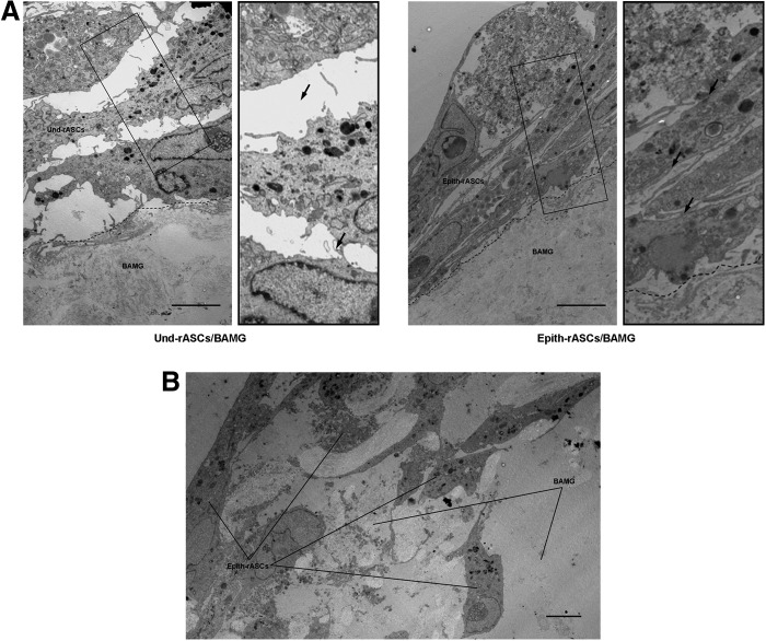
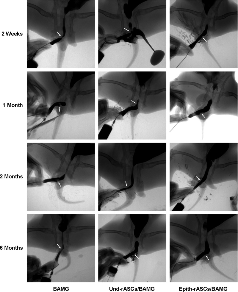
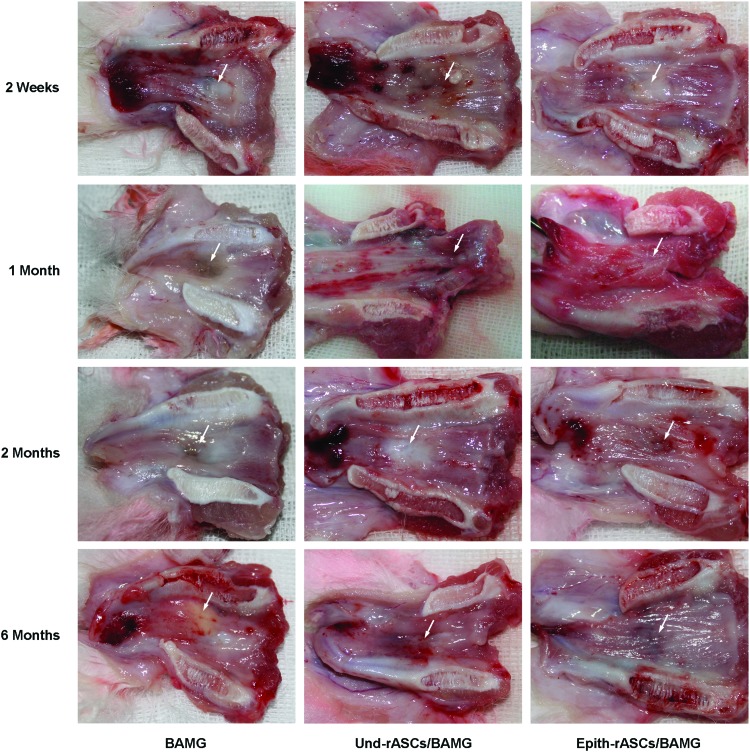
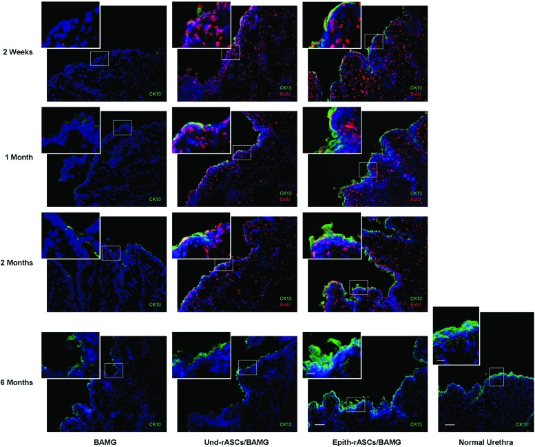
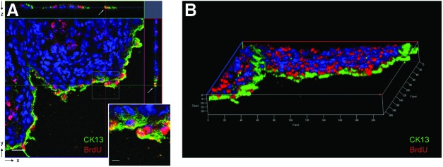
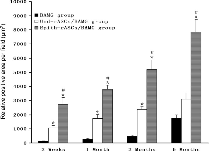
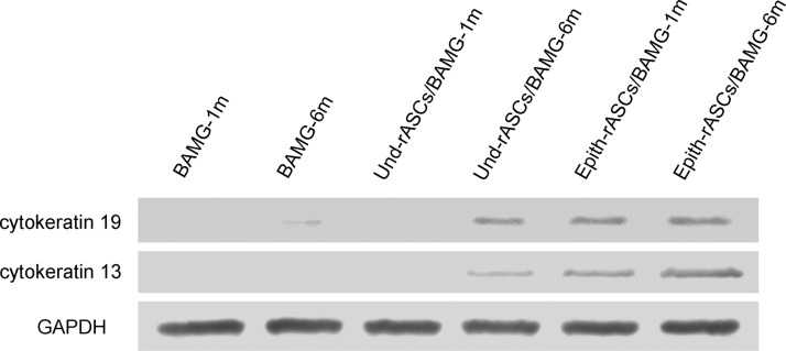
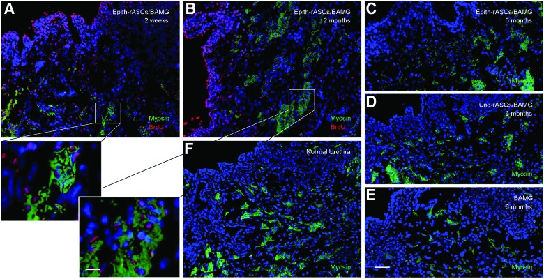
The limited amount of available epithelial tissue is considered a main cause of the high rate of urethral reconstruction failures. The aim of this study was to investigate whether epithelial-differentiated rabbit adipose-derived stem cells (Epith-rASCs) could play a role of epithelium in vivo functionally and be a potential substitute of urothelium. Substitution urethroplasty was performed to repair an anterior urethral defect in male New Zealand rabbits using Epith-rASCs seeded bladder acellular matrix grafts (BAMGs) after 5-bromo-2'-deoxyuridine (BrdU) labeling, based on the in vitro epithelial induction system we previously described. Urethroplasty with cell-free BAMGs and with undifferentiated rASCs (Und-rASCs) seeded BAMGs were performed as controls. After surgery, a notable amelioration of graft contracture and recovery of urethral continuity were observed in the Epith-rASCs/BAMG group by retrograde urethrograms and macroscopic inspection. Immunofluorescence revealed that the BrdU-labeled Epith-rASCs/Und-rASCs colocalized with cytokeratin 13 or myosin. Consistent with the results of western blotting, at early postimplantation stage, the continuous epithelial layer with local multilayered structure was observed in the Epith-rASCs/BAMG group, whereas no significant growth and local monolayer growth profile of epithelial cells were observed in the BAMG and Und-rASCs/BAMG group, respectively. The results showed that Epith-rASCs could serve as a potential substitute of urothelium for urethral tissue engineering and be available to prevent lumen contracture and subsequent complications including recurrent stricture.


Figures

<b>FIG. 1.</b>
FIG. 1.
The rabbits in the Epith-rASCs/BAMG group underwent substitution urethroplasty using Epith-rASCs seeded BAMGs. After a urethral defect with a mean length of 2.0 cm and width of 0.8 cm was created in the rabbits' ventral anterior urethra, the compound graft was placed over the defect using 6-0 vicryl sutures. Epith-rASCs, epithelial-differentiated rabbit adipose-derived stem cells; BAMG, bladder acellular matrix graft. Color images available online at www.liebertpub.com/tea
<b>FIG. 2.</b>
FIG. 2.
BrdU incorporation and cell viability assessment of Und-rASCs and Epith-rASCs after 48 h incubation with BrdU. (A) Immunofluorescent staining of the Und-rASCs and Epith-rASCs after labeling with BrdU. The Und-rASCs and Epith-rASCs (labeled with cytokeratin 19) colocalized with BrdU expression. Cytokeratin 19: green; nuclei of all the cells stained with Hoechst 33258: blue; nuclei of BrdU-labeled cells: red. Scale bars: 50 μm. (B) Proliferation of rASCs before and after labeling with BrdU, determined by DNA assay using Hoechst 33258. BrdU, 5-bromo-2′-deoxyuridine; Und-rASCs, Und-rASCs before labeling with BrdU; BrdU+ Und-rASCs, Und-rASCs after labeling with BrdU; Epith-rASCs, Epith-rASCs before labeling with BrdU; BrdU+ Epith-rASCs, Epith-rASCs after labeling with BrdU; Und-rASCs, undifferentiated rASCs. Color images available online at www.liebertpub.com/tea
<b>FIG. 3.</b>
FIG. 3.
The compound grafts were evaluated by transmission electron microscopy after 7 days of in vitro culture. (A) A stratified cellular structure was observed in both Und-rASCs seeded and Epith-rASCs seeded BAMGs. The Und-rASCs grew in a loosely organized fashion in the intercellular space, whereas the Epith-rASCs grew in a relatively compact fashion. The enlarged images of black rectangle box shown in the right side of the original figure. Arrow indicates the intercellular space. (B) The Epith-rASCs in some areas were detected to infiltrate into the underlayer of the BAMGs. Scale bars: 5 μm.
<b>FIG. 4.</b>
FIG. 4.
Retrograde urethrograms were performed for the assessment of urethral caliber at 2 weeks, 1 month, 2 months, and 6 months postimplantation. Urethral strictures were observed in both the BAMG and Und-rASCs/BAMG groups at 2 weeks and 1 month postimplantation (urethrocutaneous fistula developed in one animal in the Und-rASCs/BAMG group); at 2 months postimplantation, the stricture of the urethra was slightly improved compared with the formers. In the Epith-rASCs/BAMG group, a mild stricture of the urethra was observed at 2 weeks postimplantation, and 1 month after the surgery, the stricture was significantly improved. Arrow indicates the replaced part of urethra.
<b>FIG. 5.</b>
FIG. 5.
Macroscopic inspection of retrieved urethra at 2 weeks, 1 month, 2 months, and 6 months postimplantation. Excessive scarring and contracture of the urethral lumen at operative site was observed in the BAMG group at 2 weeks postimplantation, followed by progressive fibrosis in the next several months. Similar contracture and fibrosis was observed in the Und-rASCs/BAMG group soon after the surgery, which improved slightly by 2 months postimplantation. In the Epith-rASCs/BAMG group, a mild contracture of the urethral lumen was observed at 2 weeks postimplantation, which was markedly improved at 2 and 6 months postimplantation. Arrow indicates the replaced part of urethra. Color images available online at www.liebertpub.com/tea
<b>FIG. 6.</b>
FIG. 6.
Immunofluorescent evaluation of retrieved urethra at 2 weeks, 1 month, 2 months, and 6 months postimplantation. Normal urethra served for the comparison. In the BAMG group, almost no epithelial cells (labeled with cytokeratin 13) were observed on the urethral surface at 2 weeks postimplantation. Local discontinuous growth of epithelial cells was observed at 1 and 2 months postimplantation. In the Und-rASCs/BAMG group, occasional epithelial cell growth was observed at 2 weeks postimplantation. At 1 and 2 months postimplantation, a monolayer of epithelial cells was observed on the surface of local lumen, which colocalized with BrdU-labeled Und-rASCs. In the Epith-rASCs/BAMG group, the urethral surface was covered with continuous epithelial layer at 2 weeks postimplantation, which also colocalized with BrdU-labeled Epith-rASCs. At 1 and 2 months postimplantation, a local multilayered epithelial cells could be observed, and the multilayered structure of cells was more significant at 6 months postimplantation compared with the control groups (at 6 months postimplantation, no BrdU labeling could be detected by immunofluorescent staining). CK13: cytokeratin 13, green; nuclei of BrdU-labeled cells: red; nuclei of all the cells stained with Hoechst 33258: blue. Scale bars: 50 μm for original figures; 12.5 μm for insets. Color images available online at www.liebertpub.com/tea
<b>FIG. 7.</b>
FIG. 7.
Confocal microscopy of cytokeratin 13 expression in implanted cells in the Epith-rASCs/BAMG group. (A) The image of confocal orthogonal slices showed the colocalization of BrdU with cytokeratin 13 from x-y cross section, and the orthogonal projection of both x-z and y-z cross-sectional cuts (white arrow) at 1 month postimplantation. (B) The colocalization of BrdU with cytokeratin 13 was also shown from the 3D view of urethral tissue. CK13: cytokeratin 13, green; nuclei of BrdU labeled cells: red; nuclei of all the cells stained with Hoechst 33258: blue. Scale bars: 20 μm for original figure; 5 μm for inset. Color images available online at www.liebertpub.com/tea
<b>FIG. 8.</b>
FIG. 8.
Epithelial positive areas of cytokeratin 13 staining at each time point in various groups. *p<0.05 compared with the BAMG group; #p<0.05 compared with the Und-rASCs/BAMG group. n=5.
<b>FIG. 9.</b>
FIG. 9.
Expression of epithelial-specific markers (cytokeratin 19, cytokeratin 13) at 1 and 6 months postimplantation determined by western blot analysis. GAPDH served as the control.
<b>FIG. 10.</b>
FIG. 10.
Myosin expression in the neourethra. (A, B) BrdU immunostaining in myosin-positive cells at 2 weeks (A) and 2 months (B) postimplantation in the Epith-rASCs/BAMG group. (C–E) Immunostaining of myosin at 6 months postimplantation in the Epith-rASCs/BAMG group (C), the Und-rASCs/BAMG group (D), and the BAMG group (E), respectively. (F) Immunostaining of myosin in normal urethra. Myosin: green; nuclei of BrdU-labeled cells: red; nuclei of all the cells stained with Hoechst 33258: blue. Scale bars: 50 μm for original figures; 12.5 μm for insets. Color images available online at www.liebertpub.com/tea

References

    1. Manzoni G.Hypospadias repair failures: lessons learned. Eur Urol 49, 772, 2006 - PubMed
    1. Barbagli G., Perovic S., Djinovic R., Sansalone S., and Lazzeri M.Retrospective descriptive analysis of 1,176 patients with failed hypospadias repair. J Urol 183, 207, 2010 - PubMed
    1. Sievert K.D., Bakircioglu M.E., Nunes L., Tu R., Dahiya R., and Tanagho E.A.Homologous acellular matrix graft for urethral reconstruction in the rabbit: histological and functional evaluation. J Urol 163, 1958, 2000 - PubMed
    1. De Filippo R.E., Yoo J.J., and Atala A.Urethral replacement using cell seeded tubularized collagen matrices. J Urol 168, 1789, 2002 - PubMed
    1. Atala A.Tissue engineering for the replacement of organ function in the genitourinary system. Am J Transplant 4Suppl 6, 58, 2004 - PubMed

Publication types

MeSH terms