Review
. 2014 Jun;142(2):151-66.
doi: 10.1111/imm.12233.

Inflammation in neurodegenerative diseases--an update

Affiliations
Review

Inflammation in neurodegenerative diseases--an update

Sandra Amor et al. Immunology. 2014 Jun.

Abstract

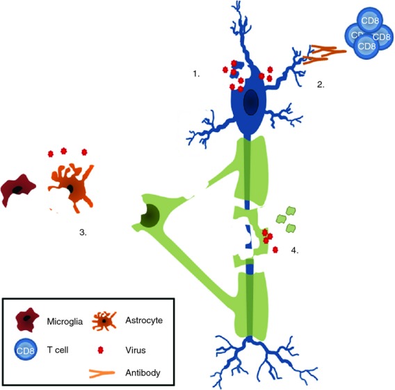
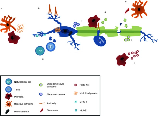
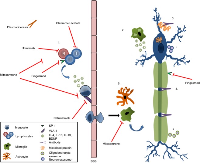
Neurodegeneration, the progressive dysfunction and loss of neurons in the central nervous system (CNS), is the major cause of cognitive and motor dysfunction. While neuronal degeneration is well-known in Alzheimer's and Parkinson's diseases, it is also observed in neurotrophic infections, traumatic brain and spinal cord injury, stroke, neoplastic disorders, prion diseases, multiple sclerosis and amyotrophic lateral sclerosis, as well as neuropsychiatric disorders and genetic disorders. A common link between these diseases is chronic activation of innate immune responses including those mediated by microglia, the resident CNS macrophages. Such activation can trigger neurotoxic pathways leading to progressive degeneration. Yet, microglia are also crucial for controlling inflammatory processes, and repair and regeneration. The adaptive immune response is implicated in neurodegenerative diseases contributing to tissue damage, but also plays important roles in resolving inflammation and mediating neuroprotection and repair. The growing awareness that the immune system is inextricably involved in mediating damage as well as regeneration and repair in neurodegenerative disorders, has prompted novel approaches to modulate the immune system, although it remains whether these approaches can be used in humans. Additional factors in humans include ageing and exposure to environmental factors such as systemic infections that provide additional clues that may be human specific and therefore difficult to translate from animal models. Nevertheless, a better understanding of how immune responses are involved in neuronal damage and regeneration, as reviewed here, will be essential to develop effective therapies to improve quality of life, and mitigate the personal, economic and social impact of these diseases.

Keywords: central nervous system; inflammation; innate; neurodegeneration; neuroprotection; repair.


Figures

Figure 1
Figure 1
Immune responses in human and experimental inflammatory neurodegenerative disorders. In an ischaemic area in stroke, HLA class II+ cells (blue, arrow) can be seen phagocytosing myelin basic protein (red) (a). In Alzheimer's disease, activated microglia (HLA class II+; blue) cluster around neurons and amyloid deposits (red) (b). A meningeal infiltrate in acute bacterial meningitis contains a single CD20+ B cell (brown; c), yet large numbers of activated microglia (HLA class II+; brown) in the meninges as well as in the brain parenchyma (d). In multiple sclerosis (MS), macrophages and microglia phagocytose myelin, and turn into so-called foam cells that are found in the perivascular space (e) as well as in the cerebrospinal fluid (f). Large numbers of inflammatory cells around a blood vessel are found in the centre of MS lesions that show extensive loss of myelin (g), as well as at the rim of the demyelinating zone (h). In the autoimmune mouse model of MS, perivascular infiltrates of HLA-class II positive cells can be observed in the white and grey matter of the spinal cord (i). In the meninges of mice that have been immunized with the neuronal antigen NF-L, the presence of B220+ B cells (j) and CD4+ T cells (k) is closely associated with neuronal damage (l).
Figure 2
Figure 2
Proposed mechanisms of viral-induced neuronal damage. Infection of neurons with viruses leads to apoptosis, necrosis, autophagy or dying back of axons (1). Immune-mediated attack of neurons by virus-specific CD8+ T cells or autoantibodies induces damage (2). Infection of endothelial cells, ependymal cells, glia (astrocytes, oligodendrocytes, microglia) leads to so-called bystander damage as the result of release of cytokines or reactive oxygen species that damage neurons in a variety of ways (see text for details) (3). Secondary neuronal degeneration as a consequence of trophic support, when oligodendrocytes and myelin are infected with viruses (4).
Figure 3
Figure 3
Immune-mediated neurodegeneration. Misfolded and aggregated proteins such as Aβ and α-synuclein aggregates (1), activate microglia and suppress brain derived neuronal growth factor production by astrocytes. Neuronal specific antibodies (2) activate the complement system or FcR-mediated damage. Natural killer and T cells damage neurons either via MHC class-I or non-classical MHC molecules (3). Damage to neurons reduces the levels of neuroimmunoregulatory molecules, decreasing the immune suppressive environment (4). Excessive production of glutamate by, for example, stressed neurons and activated immune cells, together with reduced glutamate uptake causes excitotoxic damage of neurons (5). Macrophage and microglia activation triggers release of reactive oxygen and nitrogen species, matrix metalloproteinases, chemokines and cytokines known to damage axons and neurons and to cause mitochondrial dysfunction. Neurons (i) and oligodendrocytes (ii) release exosomes that may trigger pathogenic immune responses (6). NO, nitric oxide; ROS, reactive oxygen species.
Figure 4
Figure 4
Neuroprotection and regeneration by cells of the immune system. T and B cells secrete neuroprotective factors and suppress pro-inflammatory responses (1), since macrophages/microglia that phagocytose myelin stimulate growth and repair via glial cell-derived neuronal growth factors (2). Antibodies aid remyelination thereby restoring trophic support of neurons (3) and help to remove the aggregated proteins (4). Reparative microglia and astrocytes produce neuronal growth factors (5) neurons (i) and oligodendrocytes (ii) release exosomes that dampen the pathogenic immune responses. BBB, blood–brain barrier; BDNF, brain derived neural growth factor; IL-4, interleukin-4; SP-1, sphingosine-1-phosphate; VLA-4, very-late antigen 4.

References

    1. Geneva: World Health Organization; 2012. World Health Organization Dementia a public health priority. http://site.ebrary.com/lib/ucmerced/Doc?id=10718026 [accessed 14 November 2013]
    1. Amor S, Puentes F, Baker D, et al. Inflammation in neurodegenerative diseases. Immunology. 2010;129:154–69. - PMC - PubMed
    1. Town T, Tan J, Flavell RA, et al. T-cells in Alzheimer's disease. Neuromolecular Med. 2005;7:255–64. - PubMed
    1. Bellucci A, Bugiani O, Ghetti B, et al. Presence of reactive microglia and neuroinflammatory mediators in a case of frontotemporal dementia with P301S mutation. Neurodegener Dis. 2011;8:221–9. - PMC - PubMed
    1. Mosley RL, Hutter-Saunders JA, Stone DK, et al. Inflammation and adaptive immunity in Parkinson's disease. Cold Spring Harb Perspect Med. 2012;2:a009381. - PMC - PubMed

Publication types

MeSH terms