. 2014 Apr;24(3):261-9.
doi: 10.1111/bpa.12116. Epub 2014 Feb 12.

Geniposide ameliorates learning memory deficits, reduces tau phosphorylation and decreases apoptosis via GSK3β pathway in streptozotocin-induced alzheimer rat model

Affiliations

Geniposide ameliorates learning memory deficits, reduces tau phosphorylation and decreases apoptosis via GSK3β pathway in streptozotocin-induced alzheimer rat model

Chong Gao et al. Brain Pathol. 2014 Apr.

Abstract

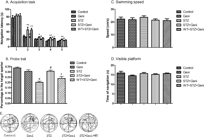
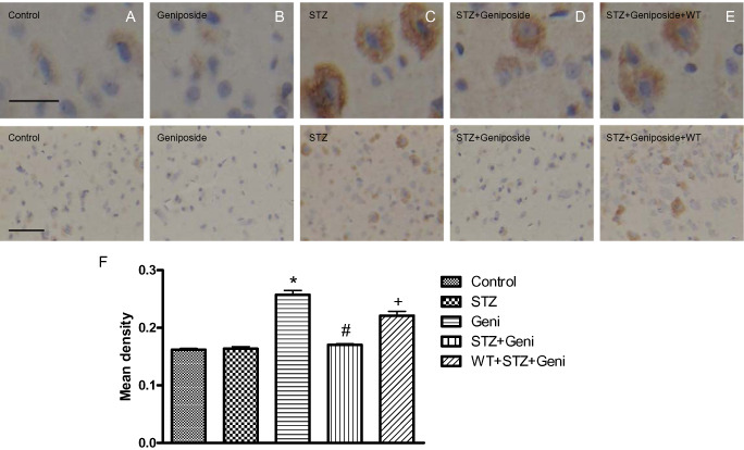
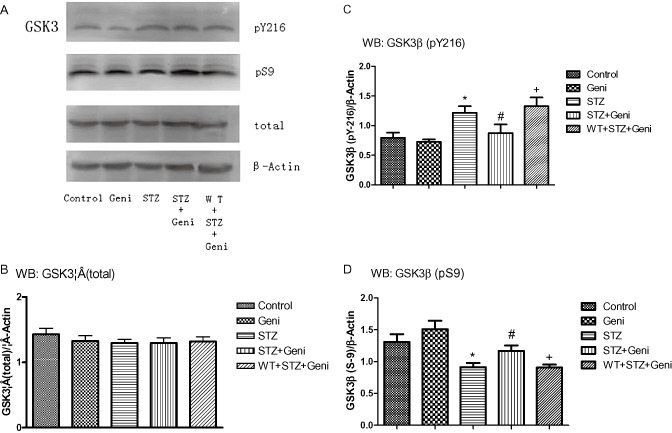
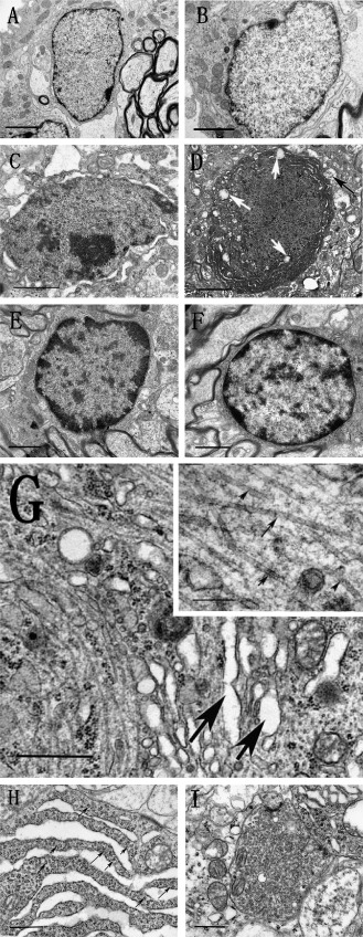
Intracerebral-ventricular (ICV) injection of streptozotocin (STZ) induces an insulin-resistant brain state that may underlie the neural pathogenesis of sporadic Alzheimer disease (AD). Our previous work showed that prior ICV treatment of glucagon-like peptide-1 (GLP-1) could prevent STZ-induced learning memory impairment and tau hyperphosphorylation in the rat brain. The Chinese herbal medicine geniposide is known to relieve symptoms of type 2 diabetes. Because geniposide is thought to act as a GLP-1 receptor agonist, we investigated the potential therapeutic effect of geniposide on STZ-induced AD model in rats. Our result showed that a single injection of geniposide (50 μM, 10 μL) to the lateral ventricle prevented STZ-induced spatial learning deficit by about 40% and reduced tau phosphorylation by about 30% with Morris water maze test and quantitative immunohistochemical analysis, respectively. It has been known that tau protein can be phosphorylated by glycogen synthase kinase-3 (GSK3) and STZ can increase the activity of GSK3β. Our result with Western blot analysis showed that central administration of geniposide resulted in an elevated expression of GSK3β(pS-9) but suppressed GSK3β(pY-216) indicating that geniposide reduced STZ-induced GSK3β hyperactivity. In addition, ultrastructure analysis showed that geniposide averted STZ-induced neural pathology, including paired helical filament (PHF)-like structures, accumulation of vesicles in synaptic terminal, abnormalities of endoplasmic reticulum (ER) and early stage of apoptosis. In summary, our study suggests that the water soluble and orally active monomer of Chinese herbal medicine geniposide may serve as a novel therapeutic agent for the treatment of sporadic AD.

Keywords: Alzheimer's disease; geniposide; glucagon-like peptide-1 receptor; glycogen synthase kinase-3β; tau hyperphosphorylation; type 2 diabetes.


Figures

Figure 1
Figure 1
Geniposide (Geni) prevented STZ (Streptozotocin)‐induced spatial learning and memory impairment. Navigation latency was significantly increased in the STZ‐treated group compared with controls on the second, third, fourth and fifth day (*, compared with control, P < 0.05; **, P < 0.01), but geniposide treatment prevented STZ‐induced learning deficit (#, compared with STZ group, P < 0.01). However, PI3K inhibitor wortmannin (WT) blocked the protective effect of geniposide (+, compared with STZ + Geniposide group, P < 0.05; ++, P < 0.01). A. STZ treatment significantly decreased the percent of time each rat spent in the target quadrant (P < 0.05), and geniposide treatment prevented STZ‐induced memory deficit. B. The probe trial did not affected by the variation of swim speed or visual acuity, respectively, of each individual rat (C,D). Video recording of navigation pattern of the five different drug treatment groups (E).
Figure 2
Figure 2
Photomicrographs and quantitative analysis of immunohistochemistry of phospho‐tau in the rat cerebral cortex. Immunoreactivity of phospho‐tau was presented as brown staining detected under higher and lower magnification, respectively (A–E, above bar = 50 μm; below bar = 150 μm). STZ treatment significantly increased phospho‐tau compared with controls (A,C,F; *, compared with control, P < 0.01), but geniposide (Geni) treatment prevented STZ‐induced increase in phospho‐tau (C,D,F; #, compared with STZ group, P < 0.01); even injection of geniposide alone did not induced the remarkable change of phosphor‐tau (A,B). PI3K inhibitor wortmannin (WT) partially prevented the protective effect of geniposide (E,F; +, compared with STZ + Geniposide group, P < 0.01).
Figure 3
Figure 3
Western blot assay of GSK3β at different phosphorylation sites in response to STZ, geniposide and WT treatment was shown in A. Total GSK3β protein was shown in the histograms of brain cortex (B). Quantitative analysis shows that no significant difference on the total expression of GSK3β among the five treatment groups (B). Phospho‐GSK3β (inactive) and (active) was presented as the ratio over loading control (β‐Actin). GSK3β activity did not differ between rats treated with aCSF and Geniposide alone (C,D). STZ alone significantly elevated GSK3β activity compared with controls as reflected with increased pGSKY216 (C, *, compared with control, P < 0.01) and decreased pGSKS9 (D, *, compared with control, P < 0.01). STZ + Geniposide prevented STZ‐induced increase in GSK3β activity, reflected as lower ratio of pGSKY216 (C, #, compared to STZ‐alone group, P < 0.01) and higher ratio of pGSKS9 (D, # compared to STZ‐alone group, P < 0.01). PI3K inhibitor WT blocked the protective effect of geniposide, reflected as higher ratio of pGSKY216 (C, + compared to STZ + Geni, P < 0.01) and lower ratio of pGSKS9 (D, + compared to STZ + Geni, P < 0.01).
Figure 4
Figure 4
Photomicrograph reveals ultrastructure alterations in the rat hippocampus under TEM in response to different drug treatment. Normal structure of hippocampal neurons that received aCSF and geniposide alone (A, B) (bar = 1 μ). Dim cells, indicative of early stages of apoptosis, that were characterized with whole cell condensation (C), dark colored mitochondria vacuolation (white arrow), and dendritic vacuolation (black arrow) (D) (bar = 1 μ) were seen in animals treated with STZ, but these signs of early apoptosis were absent in animal that were co‐administered with STZ and geniposide (E, F) (bar = 1 μ). STZ also induced the PHFs‐like structures (G, bar = 1 μ; bar = 200 nm in the higher magnification insert), which are indicative of early signs of intracellular NFTs formation. Furthermore, STZ also induced increased cleft of ER and with agglutinated ribosome (arrow) (H, bar = 1 μ), as well as elevated accumulation of neurotransmitter vesicles (I, bar = 1 μ).

References

    1. Agrawal R, Tyagi E, Shukla R, Nath C (2011) Insulin receptor signaling in rat hippocampus: a study in STZ (ICV) induced memory deficit model. Eur Neuropsychopharmacol 21:261–273. - PubMed
    1. Akter K, Lanza EA, Martin SA, Myronyuk N, Rua M, Raffa RB (2011) Diabetes mellitus and Alzheimer's disease: shared pathology and treatment? Br J Clin Pharmacol 71:365–376. - PMC - PubMed
    1. Atri A (2011) Effective pharmacological management of Alzheimer's disease. Am J Manag Care 17:S346–355. - PubMed
    1. Campion D, Dumanchin C, Hannequin D, Dubois B, Belliard S, Puel M et al (1999) Early‐onset autosomal dominant Alzheimer disease: prevalence, genetic heterogeneity, and mutation spectrum. Am J Hum Genet 65:664–670. - PMC - PubMed
    1. Carvalho C, Cardoso S, Correia SC, Santos RX, Santos MS, Baldeiras I et al (2012) Metabolic alterations induced by sucrose intake and Alzheimer's disease promote similar brain mitochondrial abnormalities. Diabetes 61:1234–1242. - PMC - PubMed

Publication types

MeSH terms