. 2014 Mar;18(3):530-41.
doi: 10.1111/jcmm.12207. Epub 2013 Dec 14.

The ROS-induced cytotoxicity of ascorbate is attenuated by hypoxia and HIF-1alpha in the NCI60 cancer cell lines

Affiliations

The ROS-induced cytotoxicity of ascorbate is attenuated by hypoxia and HIF-1alpha in the NCI60 cancer cell lines

Tobias Sinnberg et al. J Cell Mol Med. 2014 Mar.

Abstract

Intravenous application of high-dose ascorbate is used in complementary palliative medicine to treat cancer patients. Pharmacological doses of ascorbate in the mM range induce cytotoxicity in cancer cells mediated by reactive oxygen species (ROS), namely hydrogen peroxide and ascorbyl radicals. However, little is known about intrinsic or extrinsic factors modulating this ascorbate-mediated cytotoxicity. Under normoxia and hypoxia, ascorbate IC50 values were determined on the NCI60 cancer cells. The cell cycle, the influence of cobalt chloride-induced hypoxia-inducible factor-1α (HIF-1α) and the glucose transporter 1 (GLUT-1) expression (a pro-survival HIF-1α-downstream-target) were analysed after ascorbate exposure under normoxic and hypoxic conditions. The amount of ascorbyl radicals increased with rising serum concentrations. Hypoxia (0.1% O2 ) globally increased the IC50 of ascorbate in the 60 cancer cell lines from 4.5 ± 3.6 mM to 10.1 ± 5.9 mM (2.2-fold increase, P < 0.001, Mann-Whitney t-test), thus inducing cellular resistance towards ascorbate. This ascorbate resistance depended on HIF-1α-signalling, but did not correlate with cell line-specific expression of the ascorbate transporter GLUT-1. However, under normoxic and hypoxic conditions, ascorbate treatment at the individual IC50 reduced the expression of GLUT-1 in the cancer cells. Our data show a ROS-induced, HIF-1α- and O2 -dependent cytotoxicity of ascorbate on 60 different cancer cells. This suggests that for clinical application, cancer patients should additionally be oxygenized to increase the cytotoxic efficacy of ascorbate.

Keywords: GLUT-1; HIF-1α; ROS; ascorbate; cancer; hypoxia; therapy.


Figures

Figure 1
Figure 1
Ascorbate generates ascorbyl and intracellular peroxide radicals in medium and in nine different cancer cell lines. (A) Induction of Asc−• radicals from ascorbate in medium, measured by electron spin resonance (ESR) spectroscopy. Increasing serum concentrations yield more detectable Asc−• radicals. All ESR measurements were performed in triplicates; shown is one representative image for each treatment group. (B) Nine different cancer cell lines were exposed to ascorbate (8 or 16 mM) with and without the addition of 100 μg/ml catalase in full medium. The generation of H2O2 after ascorbate treatment was measured in PBS by using dichlorofluorescein substrate. We detected an increase of H2O2 in all cell lines tested; this induction of H2O2 was completely blocked by catalase. Addition of 0.5 mM H2O2 to the cell cultures served as positive control. All H2O2 measurements were performed in quadruplicates; shown is mean ± SD. (A and B) RPMI: cell culture medium, FCS: foetal calf serum, asc.: ascorbate.
Figure 2
Figure 2
Determination of the IC50 concentrations of ascorbate in the OVCAR-4 and NCI-H23 cell lines. Shown are two examples (OVCAR-4 (A), NCI-H23 (B)) of the proliferation assays and the determination of the respective IC50 values of ascorbate. The cells were exposed to 11 rising concentrations of ascorbate under normoxic (21% O2) and severe hypoxic (0.1% O2) conditions. The IC50 values were then calculated by using GraphPad Prism 5 (Nonlinear Regression EC50 shift; graphs on the right). Hypoxia increased the mean IC50 values of OVCAR-4 cells from 4.5 to 9.0 mM, and of NCI-H23 cells from 5.0 to 6.0 mM. Assays were performed in quadruplicates; shown is mean ± SEM.
Figure 3
Figure 3
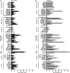
Severe hypoxia significantly increases the IC50 concentrations of ascorbate in the 60 cancer cell lines. (A) All 60 cancer cell lines of the NCI60 panel were subjected to the experimental procedures as shown in Figure 2. Shown are the calculated IC50 concentrations. (A) IC50 concentrations under normoxic conditions. (B) IC50 concentrations under hypoxic conditions. Hypoxia increased the mean IC50 value of all 60 cancer cell lines from 4.5 ± 3.6 mM to 10.1 ± 5.9 mM (2.2-fold increase, P < 0.001, Mann–Whitney t-test). Assays were performed in quadruplicates; shown is mean ± SEM.
Figure 4
Figure 4
Hypoxia-induced ascorbate resistance is driven by cobalt chloride (CoCl2)-induced hypoxia-inducible factor-1α (HIF1α) and oxygen pressure. (A and B) 100 μM CoCl2 was applied on TK10, HS578T, UACC257, HT29, SNB19 and OVCAR8 cells under normoxic and hypoxic conditions for 24 hrs. The Western blot and densitometric analyses show a strong induction of HIF1α upon CoCl2-treatment in five of the six cell lines (C). The influence of hypoxia alone, CoCl2-induced HIF1α or both on cancer cell viability after ascorbate exposure at increasing concentrations (0, 4, 8, and 16 mM) for 1 hr was analysed. Hypoxia induced an ascorbate resistance in all of the cells. The same was observed for CoCl2-treatment under normoxic conditions in TK10, HS578T, UACC257 and HT29 cells. Their combination (hypoxia + CoCl2-treatment) significantly increased the ascorbate resistance in all cells. The addition of catalase blocked the ascorbate-induced proliferation inhibition. N: normoxia (21% O2), H: hypoxia (0.1% O2). *P < 0.05, **P < 0.01, ***P < 0.001; ****P < 0.0001 (Two-way anova). Assays were performed in sixtuplicates; shown is mean ± SD.
Figure 5
Figure 5
Hypoxia and high-dose ascorbate increase the sub-G1 cell population in the 60 cancer cell lines. FACS cell-cycle analyses (100,000 cells measured per treatment group) were performed on all 60 cancer cell lines after incubation with or without ascorbate at the individual IC50 concentration for 24 hrs under normoxic (21% O2) and hypoxic (0.1% O2) conditions. Depicted is an exemplary cell line of each of the nine tumour entities of the NCI60 panel. The entire data are displayed in Table S2, Ascorbate treatment induced an increase in the sub-G1 cell population (apoptosis) under normoxic and hypoxic conditions. N: normoxia (21% O2), H: hypoxia (0.1% O2).
Figure 6
Figure 6
Hypoxia and ascorbate treatment alter the expression of glucose transporter 1 (GLUT-1) in the 60 cancer cell lines. Western blot analyses were performed on the cell lysates of all 60 cell lines 24 hrs after exposure to ascorbate at the individual IC50 concentration for 1 hr under normoxic and hypoxic conditions. Protein expression was evaluated by densitometric analyses. Under hypoxia, the GLUT-1 protein level increased in 40 of the cell lines, most pronounced in melanoma and breast cancer cells. Ten cell lines had a reduced GLUT-1 expression under hypoxic conditions (e.g. in the non-small cell lung cancer cells). Under normoxia, ascorbate treatment reduced GLUT-1 expression in 30 cell lines, most pronounced in melanoma, non-small cell lung cancer and breast cancer cells; 22 cell lines showed an increased GLUT-1 expression, most pronounced in ovarian and leukaemia cancer cells. Under hypoxia + high-dose ascorbate, 20 cell lines had a reduced GLUT-1 expression, which was most pronounced in the breast cancer cells, while 31 cell lines had an increased GLUT-1 expression, most pronounced in ovarian and colon cancer cells.

References

    1. Cameron E, Pauling L. Supplemental ascorbate in the supportive treatment of cancer: prolongation of survival times in terminal human cancer. Proc Natl Acad Sci USA. 1976;73:3685–9. - PMC - PubMed
    1. Padayatty SJ, Levine M. Reevaluation of ascorbate in cancer treatment: emerging evidence, open minds and serendipity. J Am Coll Nutr. 2000;19:423–5. - PubMed
    1. Chen Q, Espey MG, Krishna MC, et al. Pharmacologic ascorbic acid concentrations selectively kill cancer cells: action as a pro-drug to deliver hydrogen peroxide to tissues. Proc Natl Acad Sci USA. 2005;102:13604–9. - PMC - PubMed
    1. Chen Q, Espey MG, Sun AY, et al. Pharmacologic doses of ascorbate act as a prooxidant and decrease growth of aggressive tumor xenografts in mice. Proc Natl Acad Sci USA. 2008;105:11105–9. - PMC - PubMed
    1. Du J, Martin SM, Levine M, et al. Mechanisms of ascorbate-induced cytotoxicity in pancreatic cancer. Clin Cancer Res. 2010;16:509–20. - PMC - PubMed

Publication types

MeSH terms