. 2013 Dec 12:10:152.
doi: 10.1186/1742-4690-10-152.

CD8+ T cells from HLA-B*57 elite suppressors effectively suppress replication of HIV-1 escape mutants

Affiliations

CD8+ T cells from HLA-B*57 elite suppressors effectively suppress replication of HIV-1 escape mutants

Christopher W Pohlmeyer et al. Retrovirology. .

Abstract

Background: Elite Controllers or Suppressors (ES) are HIV-1 positive individuals who maintain plasma viral loads below the limit of detection of standard clinical assays without antiretroviral therapy. Multiple lines of evidence suggest that the control of viral replication in these patients is due to a strong and specific cytotoxic T lymphocyte (CTL) response. The ability of CD8+ T cells to control HIV-1 replication is believed to be impaired by the development of escape mutations. Surprisingly, viruses amplified from the plasma of ES have been shown to contain multiple escape mutations, and it is not clear how immunologic control is maintained in the face of virologic escape.

Results: We investigated the effect of escape mutations within HLA*B-57-restricted Gag epitopes on the CD8+ T cell mediated suppression of HIV-1 replication. Using site directed mutagenesis, we constructed six NL4-3 based viruses with canonical escape mutations in one to three HLA*B-57-restricted Gag epitopes. Interestingly, similar levels of CTL-mediated suppression of replication in autologous primary CD4+ T cells were observed for all of the escape mutants. Intracellular cytokine staining was performed in order to determine the mechanisms involved in the suppression of the escape variants. While low baseline CD8+ T cells responses to wild type and escape variant peptides were seen, stimulation of PBMC with either wild type or escape variant peptides resulted in increased IFN-γ and perforin expression.

Conclusions: These data presented demonstrate that CD8+ T cells from ES are capable of suppressing replication of virus harboring escape mutations in HLA-B*57-restricted Gag epitopes. Additionally, our data suggest that ES CD8+ T cells are capable of generating effective de novo responses to escape mutants.


Figures
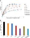
Figure 1
Figure 1
Fitness cost of canonical HLA-B*57 escape mutations. A. Average infection by seven NL4-3 escape variants in uninfected individuals. Viral inoculum was quantified by relative qPCR. 105 CD4+ T cells were infected in a 96-well plate in triplicate. Infection was determined by GFP expression by flow cytometry. Wild type NL4-3 (black) showed the highest level of infection, while NL4-3 escape mutant variants showed reduced maximal infection (I147L, orange; A146P, navy; A163S, red; A146P/A163S, purple; T242N/G248A, brown; A146P/A163S/T242N/G248A, teal). Error bars represent SEM. n = 4. B. Maximal infection of each escape variant is compared relative to wild type NL4-3 virus.
Figure 2
Figure 2
Suppression of replication of NL4-3 escape variant viruses. A-C: Unstimulated CD4+ T cells from HLA-B*5703 positive ES were infected with one of seven NL4-3 variants (wild type, black; I147L, orange; A146P, navy; A163S, red; A146P/A163S, purple; T242N/G248A, brown; A146P/A163S/T242N/G248A, teal) and cultured with autologous CD8+ T cells isolated from fresh PBMCs (A) or PBMCs stimulated with either wild type (B) or escape variant (C) HLA-B*57- Gag restricted Gag peptides for 7 days before isolation. CD8+ T cells were co-cultured with infected CD4+ T cells at three effector to target ratios. D-F: Unstimulated CD4+ T cells from healthy donors were infected with one of the seven NL4-3 variants used above and co-cultured with CD8+ T cells as was done with ES (D, unstimulated; E, wild type peptide stimulated; F, escape variant stimulated). Infection of CD4+ T cells was quantified by flow cytometry on days 3 (left) and 5 (right) after infection by GFP expression. Median values are plotted. For ES, n = 7. For healthy donors, n = 5.
Figure 3
Figure 3
Individual suppression of NL4-3 escape variant viruses. Unstimulated CD4+ T cells from HLA-B*5703+ ES were infected with one of seven NL4-3 variants (wild type, black; I147L, orange; A146P, navy; A163S, red; A146P/A163S, purple; T242N/G248A, brown; A146P/A163S/T242N/G248A, teal) and cultured with autologous unstimulated CD8+ cells at three effector to target ratios. A shows maximal suppression on day 3; B shows maximal suppression on day 5. Black horizontal bars indicate median. Asterisk indicates P < 0.05.
Figure 4
Figure 4
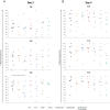
Intracellular cytokine staining of ES CD8+ T cells. A-D: CD8+ T cells of HLA-B*57 positive ES were either freshly isolated (left) or stimulated with either wild type (center) or escape variant (right) HLA-B*57-restricted Gag peptides for 7 days. Cells from each group underwent an overnight stimulation with individual peptides. Percentage of CD8+ T cells expressing IFN-γ when stimulated overnight with TW10 (A), KF11 (B), and IW9 (C) in blue, or the escape mutant variant peptide containing T242N/G248A (A), A163S (B), and I147L (C) mutations in red is shown. E-H: Percentage of CD8+ T cells expressing both IFN-γ and perforin after restimulation with TW10 (E), KF11 (F), and IW9 (B) in blue, or the escape mutant variant peptide containing T242N/G248A (E), A163S (F), and I147L (G) in red is shown. D and H show CD8+ T cells that express IFN-γ or co-express IFN-γ and perforin when PBMCs were stimulated overnight with Gag 263-272 (KK10, HLA-B*27+ peptide). Black horizontal bars indicate statistically significant difference (P < 0.05) between samples when stimulated overnight with the same variant peptide; gray horizontal bars indicate statistically significant difference (P < 0.05) between samples when stimulated overnight with opposite variant peptide.
Figure 5
Figure 5
Intracellular cytokine staining of CP CD8+ T cells. A-D: CD8+ T cells of HLA-B*57 positive CP were either freshly isolated (left) or stimulated with either wild type (center) or escape variant (right) HLA-B*57-restricted Gag peptides for 7 days. CPs were either on suppressive HAART regiments with undetectable viral loads (circles), were on HAART regimens but recently had detectable levels of viremia (triangles), or were not on HAART and had high levels of viremia (diamond). Cells from each group underwent an overnight stimulation with individual peptides. Percentage of CD8+ T cells expressing IFN-γ when restimulated with TW10 (A), KF11 (B), and IW9 (C) in blue, or the escape mutant variant peptide containing T242N/G248A (A), A163S (B), and I147L (C) mutations in red is shown. E-H: Percentage of CD8+ T cells expressing both IFN-γ and perforin after overnight stimulation with TW10 (E), KF11 (F), and IW9 (G) in blue, or the escape mutant variant peptide containing T242N/G248A (E), A163S (F), and I147L (G) mutations in red is shown. D and H show CD8+ T cells that express IFN-γ or co-express IFN-γ and perforin when PBMCs were stimulated overnight with Gag 263-272 (KK10, HLA-B*27+ peptide). Black asterisks indicate statistically significant difference (P < 0.05) between samples when restimulated with the same variant peptide; gray asterisks indicates statistically significant difference (P < 0.05) between samples when restimulated with opposite variant peptide.

Similar articles

Cited by

References

    1. Okulicz JF, Lambotte O. Epidemiology and clinical characteristics of elite controllers. Curr Opin HIV AIDS. 2011;10(3):163–168. doi: 10.1097/COH.0b013e328344f35e. - DOI - PubMed
    1. O’Connell K a, Bailey JR, Blankson JN. Elucidating the elite: mechanisms of control in HIV-1 infection. Trends Pharmacol Sci. 2009;10:631–637. doi: 10.1016/j.tips.2009.09.005. - DOI - PubMed
    1. Deeks SG, Walker BD. Human immunodeficiency virus controllers: Mechanisms of durable virus control in the absence of antiretroviral therapy. Immunity. 2007;10:406–416. doi: 10.1016/j.immuni.2007.08.010. - DOI - PubMed
    1. Migueles SA, Connors M. Long-term nonprogressive disease among untreated HIV-infected individuals: Clinical implications of understanding immune control of HIV. JAMA. 2010;10:194–201. doi: 10.1001/jama.2010.925. - DOI - PubMed
    1. Deacon NJ, Tsykin A, Solomon A, Smith K, Ludford-Mending M, Hooker DJ, McPhee DA, Greenway AL, Ellett A, Chatfield C, Lawson VA, Crowe S, Maerz A, Sonza S, Learmont J, Sullivan JS, Cunningham A, Dwyer D, Dowton D, Mills J. Genomic structure of an attenuated quasi species of HIV-1 from a blood transfusion donor and recipients. Science. 1995;10:988–991. doi: 10.1126/science.270.5238.988. - DOI - PubMed

Publication types

LinkOut - more resources