. 2014 Feb;133(2):222-8.
doi: 10.1016/j.thromres.2013.11.009. Epub 2013 Nov 16.

Anti-human protein S antibody induces tissue factor expression through a direct interaction with platelet phosphofructokinase

Affiliations

Anti-human protein S antibody induces tissue factor expression through a direct interaction with platelet phosphofructokinase

Changyi Chen et al. Thromb Res. 2014 Feb.

Abstract

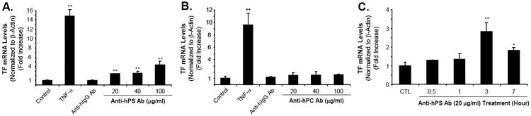
Introduction: Autoantibodies including anti-human protein S antibody (anti-hPS Ab) and anti-human protein C antibody (anti-hPC Ab) can be detected in patients with autoimmune diseases with hypercoagulability. The objective of the present study was to determine the effects and molecular pathways of these autoantibodies on tissue factor (TF) expression in human coronary artery endothelial cells (HCAECs).

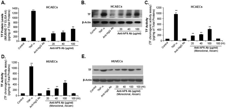
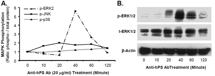
Materials and methods: HCAECs were treated with anti-hPS Ab or anti-hPC Ab for 3 hours. TF expression was measured by real-time PCR and Western blot. TF-mediated procoagulant activity was determined by a commercial kit. MAPK phosphorylation was analyzed by Bio-Plex luminex immunoassay and Western blot. The potential proteins interacting with anti-hPS Ab were studied by immunoprecipitation, mass spectrometry and in vitro pull-down assay.

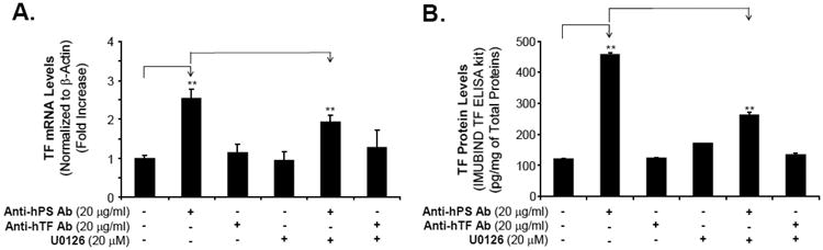
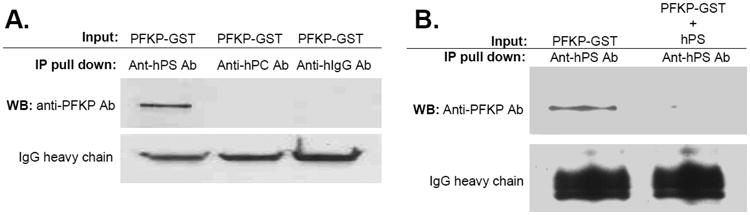
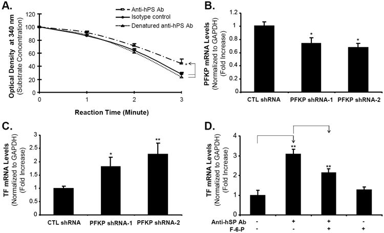
Results: Anti-hPS Ab, but not anti-hPC Ab, specifically induced TF expression and TF-mediated procoagulant activity in HCAECs in a concentration-dependent manner. This effect was confirmed in human umbilical endothelial cells (HUVECs). ERK1/2 phosphorylation was induced by anti-hPS Ab treatment, while inhibition of ERK1/2 by U0216 partially blocked anti-hPS Ab-induced TF upregulation (P<0.05). In addition, anti-hPS Ab specifically cross-interacted with platelet phosphofructokinase (PFKP) in HCAECs. Anti-hPS Ab was able to directly inhibit PFKP activities in HCAECs. Furthermore, silencing of PFKP by PFKP shRNA resulted in TF upregulation in HCAECs, while activation of PFKP by fructose-6-phosphate partially blocked the effect of anti-hPS Ab on TF upregulation (P<0.05).

Conclusions: Anti-hPS Ab induces TF expression through a direct interaction with PFKP and ERK1/2 activation in HCAECs. Anti-hPS Ab may directly contribute to vascular thrombosis in the patient with autoimmune disorders.

Keywords: Anti-human protein S antibody; Autoantibody; ERK1/2; Endothelial cell; Platelet phosphofructokinase; Tissue factor.


Conflict of interest statement

Conflict of Interest Statement: None

Figures

Fig. 1
Fig. 1
Effect of anti-hPS Ab on TF mRNA levels in HCAECs. (A). Concentration-dependent response of anti-hPS Ab (polyclonal, Sigma) in TF mRNA expression. (B). Concentration-dependent response of anti-hPC Ab in TF mRNA expression. HCAECs were serum-starved for 12-16 hours, and then treated with 20-100 μg/mL anti-hPS Ab or anti-hPC Ab, 100 μg/mL anti-hIgG Ab or 5 ng/mL TNF-α for 3 hours. (C). Time course of anti-hPS Ab-induced TF mRNA expression. HCAECs were serum-starved for 12-16 hours, and then treated with 20 μg/mL anti-hPS Ab for indicated time points. TF mRNA expression was measured by real-time PCR. The relative amount of TF mRNA was normalized to β-actin. Data shown are the mean + SEM of triplicate determinations. **P < 0.01 versus untreated controls.
Fig. 2
Fig. 2
Effect of anti-hPS Ab on TF protein levels as well as TF-mediated procoagulant activity in HCAECs and HUVECs. (A). Concentration-dependent response of anti-hPS Ab-induced TF protein levels (ELISA) in HCAECs. Serum-starved HCAECs were treated with 20-100 μg/mL anti-hPS Ab (polyclonal, Sigma), 100 μg/mL anti-hIgG Ab or 5 ng/mL TNF-a for 3 hours. The cellular TF protein levels were measured by IMUBIND Tissue Factor ELISA Kit. (B). Concentration dependent response of anti-hPS Ab-induced TF protein levels (Western blot) in HCAECs. Serum-starved HCAECs were treated with 20-100 μg/mL anti-hPS Ab, 100 μg/mL anti-hIgG Ab or 5 ng/mL TNF-α for 3 hours. TF protein expression was detected by Western blot. β-actin was used as a loading control. (C). Effect of anti-hPS Ab (monoclonal, Abcam) on TF-mediated procoagulant activity in HCAECs. Serum-starved HCAECs were treated with 20-100 μg/mL anti-hPS Ab, 40 μg/mL anti-hIgG Ab, 5 ng/mL TNF-a or heat-inactivated anti-hPS Ab (100 ng/mL) for 3 hours. The TF activity levels were measured by a tissue factor human chromogenic activity assay kit (Abcam). (D). Effect of anti-hPS Ab (monoclonal, Abcam) on TF-mediated procoagulant activity in HUVECs. (E). Effect of anti-hPS Ab (monoclonal, Abcam) on TF expression (Western blot) in HUVECs. Data shown are the mean + SEM of triplicate determinations. **P < 0.01 versus untreated controls.
Fig. 3
Fig. 3
Effect of anti-hPS Ab on MAPK phosphorylation in HCAECs. (A). The activation status of MAPKs (ERK2, JNK and p38) was analyzed by Bio-Plex immunoassay. Serum-starved HCAECs were treated with anti-hPS Ab (20 μg/mL) and the cell lysates were harvested at different time points with Bio-Plex Cell Lysis Kit. The phosphoprotein and total proteins of MAPKs were analyzed by a Luminex 100TM analyzer and Bio-Plex Manager software (BioRad). (B). Phosphorylation of ERK1/2 was detected by Western blot. Serum-starved HCAECs were treated with anti-PS Ab (20 μg/mL) for different time points. The phosphorylated and total ERK1/2 proteins were detected by Western blot. β-actin was used as a loading control. Representative results from 3 experiments are shown.
Fig. 4
Fig. 4
Effect of ERK1/2 inhibitor on anti-hPS Ab-induced TF expression and activity in HCAECs. (A). Effect of ERK1/2 inhibitor on anti-hPS Ab-induced TF mRNA expression in HCAECs. Serum-starved HCAECs we incubated with anti-hPS Ab or anti-hTF Ab, with the presence or absence of ERK1/2 inhibitor (U0126) for 3 hours. TF mRNA expression was measured by real-time PCR (BioRad). The relative amount of TF mRNA was normalized to β-actin. Data shown are the mean + SEM of triplicate determinations. (B). Effect of MEK1/2 inhibitor on anti-hPS Ab-induced TF protein activity in HCAECs. Serum-starved HCAECs were incubated with anti-hPS Ab or anti-hTF Ab, with the presence or absence of ERK1/2 inhibitor (U0126) for 3 hours. The cellular TF activity was measured by IMUBIND Tissue Factor ELISA Kit. Data shown are the mean + SEM of duplicate determinations. **P < 0.01 versus untreated controls. #P < 0.01 versus anti-hPS Ab group.
Fig. 5
Fig. 5
Specific interaction between anti-hPS Ab and PFKP. (A). Immunoprecipitation assay. Purified recombinant human PFKP-GST (0.5 μg) was mixed with anti-hPS Ab (2 μg) or control Abs (anti-hPC Ab and anti-hIgG Ab) (2 μg) at 4°C for 2 hours, followed by incubation with protein A agarose beads for additional 1 hour. Bound proteins were subjected to SDS-PAGE and immunoblotted with anti-PFKP Ab. (B). Human protein S abolished the binding activity between PFKP-GST and anti-hPS Ab. Purified recombinant human PFKP-GST (0.5 μg) was mixed with anti-hPS Ab (2 μg) and human protein S (20 μg) at 4°C for 2 hours, followed by incubation with protein A agarose beads for additional 1 hour. Bound proteins were subjected to SDS-PAGE and immunoblotted with anti-PFKP Ab. Representative results from 3 experiments are shown.
Fig. 6
Fig. 6
Role of PFKP in anti-hPS Ab-induced TF expression in HCAECs. (A). PFKP activity assay. The PFKP crude extract from HCAECs was incubated with anti-hPS Ab, heat denatured-anti-hPS Ab or isotype control Ab for 15 minutes, and then the PFKP activity was analyzed in the reaction mixture for 3 minutes at room temperature. The PFKP activity was represented as percent loss of DPNH in the reaction at A340. (B). PFKP mRNA expression in stably transfected HCAECs. The 29mer shRNA constructs against PFKP and control construct plasmid were transfected into Phoenix Ampho packaging cells to produce recombinant retroviruses. HCAECs were then infected by the viral supernatants and selected in growth medium containing 1 μg/mL puromycin for 1 week. The PFKP mRNA expression in stably transfected HCAECs was measured by real-time PCR. (C). TF mRNA expression in stably transfected HCAECs. The TF mRNA expression in stably transfected HCAECs was measured by real-time PCR. (D). Fructose-6-phosphate blocked the effects of anti-hPS Ab. Serum-starved HCAECs were incubated with anti-hPS Ab in the presence or absence of PFKP activator (fructose-6-phosphate) for 3 hours. TF mRNA expression was measured by real-time PCR. The relative amount of specific gene mRNA was normalized to GAPDH. Data shown are the mean + SEM of triplicate determinations. *P < 0.05 and **P < 0.01 versus untreated controls. #P < 0.01 versus anti-hPS Ab group.

References

    1. Walker FJ. Protein S and the regulation of activated protein C. Semin Thromb Hemost. 1984;10:131–8. - PubMed
    1. Bereczky Z, Kovács KB, Muszbek L. Protein C and protein S deficiencies: similarities and differences between two brothers playing in the same game. Clin Chem Lab Med. 2010;48(Suppl 1):S53–66. - PubMed
    1. D'Angelo A, Della Valle P, Crippa L, Pattarini E, Grimaldi LM, Vigano D'Angelo S. Brief report: autoimmune protein S deficiency in a boy with severe thromboembolic disease. N Engl J Med. 1993;328:1753–7. - PubMed
    1. Walker FJ. Regulation of activated protein C by protein S. The role of phospholipid in factor Va inactivation. J Biol Chem. 1981;256:11128–31. - PubMed
    1. Suh CH, Hilliard B, Li S, Merrill JT, Cohen PL. TAM receptor ligands in lupus: protein S but not Gas6 levels reflect disease activity in systemic lupus erythematosus. Arthritis Res Ther. 2010;12:R146. - PMC - PubMed

Publication types

MeSH terms