Cardiovascular manifestations of heterotaxy and related situs abnormalities assessed with CT angiography
- PMID: 24331937
- PMCID: PMC3947807
- DOI: 10.1016/j.jcct.2013.11.008
Cardiovascular manifestations of heterotaxy and related situs abnormalities assessed with CT angiography
Abstract
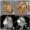
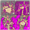
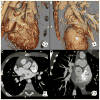
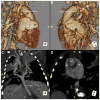
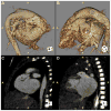
Heterotaxy and situs abnormalities describe an abnormal arrangement of visceral organs in the thoracoabdominal cavity across the normal left-right axis of the body. It is associated with a high occurrence of congenital heart and abdominal defects, including anomalous pulmonary venous connections, systemic venous abnormalities, asplenia, and intestinal malrotation. Without proper diagnosis and surgical intervention, the prognosis of patients with heterotaxy syndrome and associated congenital defects is extremely poor. Complex intracardiac and extracardiac lesions are common in heterotaxy and can be difficult to assess by echocardiography. CT angiography (CTA) is a useful tool in this setting to accurately assess intracardiac and extracardiac abnormalities in this population for medical or surgical management. The intention of this pictorial essay is to review the most common cardiovascular defects involved with heterotaxy syndrome in addition to emphasizing the utility of CTA in the identification and classification of anomalies seen in these patients. This review briefly defines most common terminology used in situs abnormalities as well as presents CT images and 3-dimensional reconstructions of common anomalies associated with situs abnormalities. In summary, this review should prepare radiologists and pediatric cardiologists to describe heterotaxy and situs abnormalities in addition to recognizing the utility of CTA in these patients.
Keywords: CT angiography; Heterotaxy; Isomerism; Situs abnormalities; Situs ambiguus; Situs inversus.
Copyright © 2013 Society of Cardiovascular Computed Tomography. Published by Elsevier Inc. All rights reserved.
Conflict of interest statement
Figures



Similar articles
-
The nomenclature, definition and classification of cardiac structures in the setting of heterotaxy.Cardiol Young. 2007 Sep;17 Suppl 2:1-28. doi: 10.1017/S1047951107001138. Cardiol Young. 2007. PMID: 18039396 Review.
-
Characterisation of computed tomography angiography findings in paediatric patients with heterotaxy.Pediatr Radiol. 2019 Aug;49(9):1142-1151. doi: 10.1007/s00247-019-04434-0. Epub 2019 Jun 5. Pediatr Radiol. 2019. PMID: 31165901
-
Human Laterality Disorders: Pathogenesis, Clinical Manifestations, Diagnosis, and Management.Am J Med Sci. 2021 Sep;362(3):233-242. doi: 10.1016/j.amjms.2021.05.020. Epub 2021 May 28. Am J Med Sci. 2021. PMID: 34052215 Review.
-
Disharmonious Patterns of Heterotaxy and Isomerism: How Often Are the Classic Patterns Breached?Circ Cardiovasc Imaging. 2018 Feb;11(2):e006917. doi: 10.1161/CIRCIMAGING.117.006917. Circ Cardiovasc Imaging. 2018. PMID: 29444810
-
Rare Extracardiac Anomalies Presented with Right Heterotaxy Syndrome in a Newborn Baby: A Case Report.Am J Case Rep. 2020 Jun 3;21:e923341. doi: 10.12659/AJCR.923341. Am J Case Rep. 2020. PMID: 32491997 Free PMC article.
Cited by
-
Heterotaxy polysplenia syndrome in an adult female with complete endocardial cushion defect.Radiol Case Rep. 2021 Feb 24;16(5):1080-1084. doi: 10.1016/j.radcr.2021.02.015. eCollection 2021 May. Radiol Case Rep. 2021. PMID: 33717387 Free PMC article.
-
Repair of a juxtarenal abdominal aortic aneurysm in a patient with situs inversus totalis using a retroperitoneal approach.J Vasc Surg Cases Innov Tech. 2016 Aug 20;2(3):92-94. doi: 10.1016/j.jvscit.2016.04.002. eCollection 2016 Sep. J Vasc Surg Cases Innov Tech. 2016. PMID: 38827209 Free PMC article.
-
Heterotaxy Syndrome With Right Isomerism and Interrupted Inferior Vena Cava: A Case Report and Literature Review.Cureus. 2024 Mar 7;16(3):e55698. doi: 10.7759/cureus.55698. eCollection 2024 Mar. Cureus. 2024. PMID: 38586636 Free PMC article.
-
Mitochondrial Dysfunction in Congenital Heart Disease.J Cardiovasc Dev Dis. 2025 Jan 25;12(2):42. doi: 10.3390/jcdd12020042. J Cardiovasc Dev Dis. 2025. PMID: 39997476 Free PMC article. Review.
-
Atrioventricular Discordance with Double-Outlet Right Ventricle in Mirror Imagery and Levocardia: A Very Rare Case Report.J Cardiovasc Echogr. 2020 Oct-Dec;30(4):227-230. doi: 10.4103/jcecho.jcecho_65_20. Epub 2021 Jan 20. J Cardiovasc Echogr. 2020. PMID: 33828947 Free PMC article.
References
-
- Balan A, Lazoura O, Padley SP, Rubens M, Nicol ED. Atrial isomerism: a pictorial review. J Cardiovasc Comput Tomogr. 2012;6:127–136. - PubMed
-
- Tonkin IL, Tonkin AK. Visceroatrial situs abnormalities: sonographic and computed tomographic appearance. AJR Am J Roentgenol. 1982;138:509–515. - PubMed
-
- Jacobs JP, Anderson RH, Weinberg PM, et al. The nomenclature, definition and classification of cardiac structures in the setting of heterotaxy. Cardiol Young. 2007;17(suppl 2):1–28. - PubMed
-
- Hagler DJ, O’Leary PW. Cardiac malpositions and abnormalities of atrial and visceral situs. In: Emmanouilides GC, Riemenschneider TA, Allen HD, Gutgesell HP, editors. Moss and Adams’ Heart Disease in Infants, Children, and Adolescents: Including the Fetus and Young Adult. Vol. 2. Baltimore, MD: Williams & Wilkins; 1995. pp. 1307–1336.
Publication types
MeSH terms
Grants and funding
LinkOut - more resources
Full Text Sources
Other Literature Sources
Medical

