. 2013 Dec 9;24(6):751-65.
doi: 10.1016/j.ccr.2013.10.013.

Transformation of the fallopian tube secretory epithelium leads to high-grade serous ovarian cancer in Brca;Tp53;Pten models

Affiliations

Transformation of the fallopian tube secretory epithelium leads to high-grade serous ovarian cancer in Brca;Tp53;Pten models

Ruth Perets et al. Cancer Cell. .

Abstract

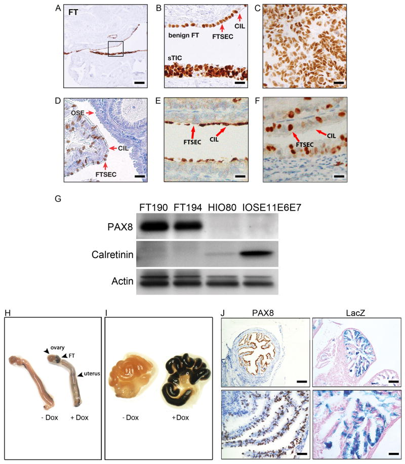
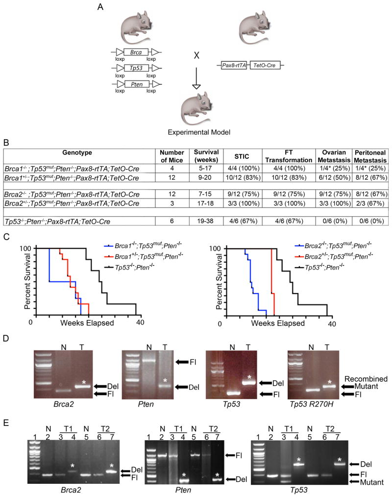
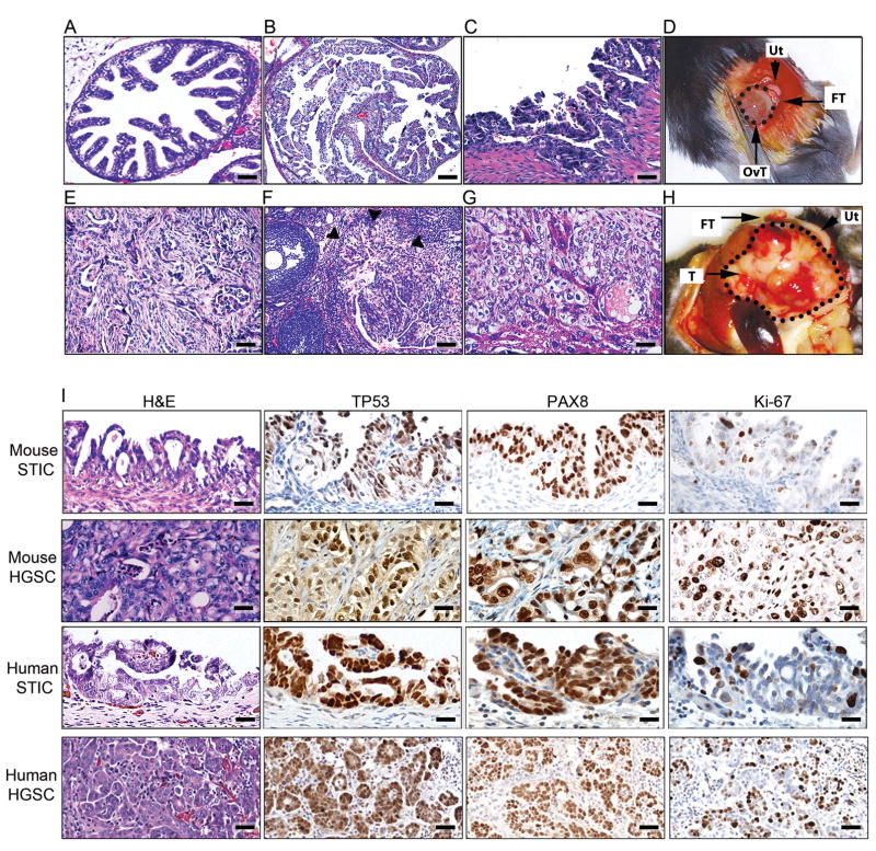
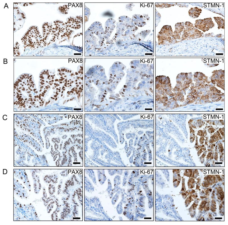
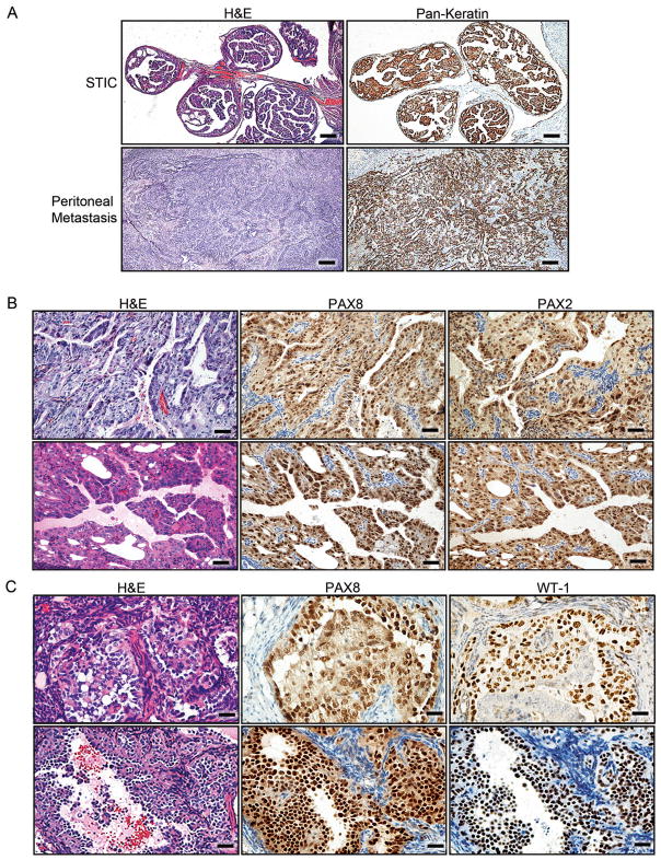
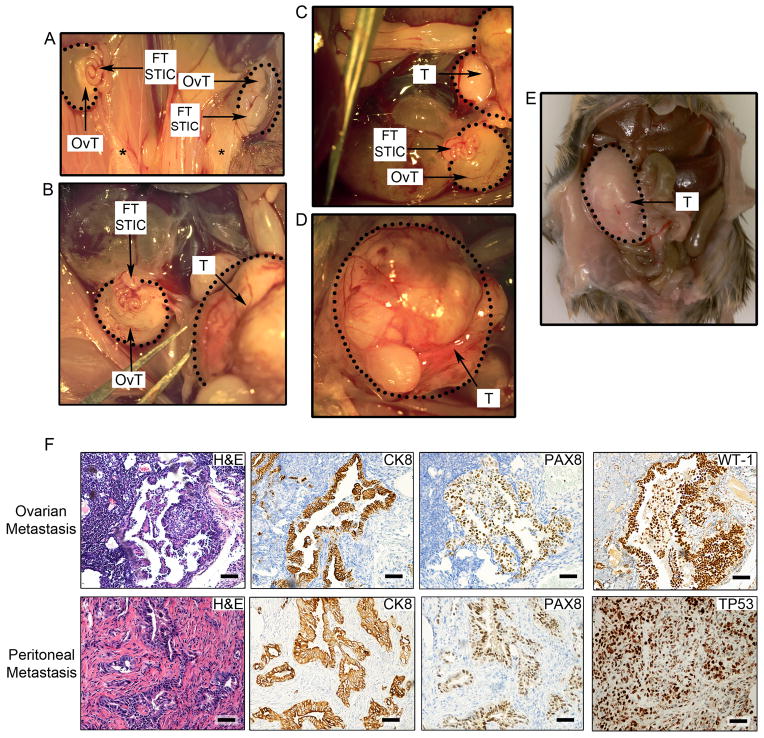
High-grade serous ovarian carcinoma presents significant clinical and therapeutic challenges. Although the traditional model of carcinogenesis has focused on the ovary as a tumor initiation site, recent studies suggest that there may be additional sites of origin outside the ovary, namely the secretory cells of the fallopian tube. Our study demonstrates that high-grade serous tumors can originate in fallopian tubal secretory epithelial cells and also establishes serous tubal intraepithelial carcinoma as the precursor lesion to high-grade serous ovarian and peritoneal carcinomas in animal models targeting the Brca, Tp53, and Pten genes. These findings offer an avenue to address clinically important questions that are critical for cancer prevention and early detection in women carrying BRCA1 and BRCA2 mutations.


Conflict of interest statement

Competing Interests Statement

The authors declare that they have no competing financial interests.

Figures

Figure 1
Figure 1. Expression of PAX8, a specific Müllerian lineage marker, during FT malignant transformation
(A, B) Immunohistochemistry (IHC) for PAX8 in the human FTE is shown for the benign epithelium (top) and STIC (bottom). The square area in (A) is shown at a higher magnification in (B). PAX8 positive FTSEC and PAX8-negative ciliated cells (CIL) are demarcated by red arrows. (C) IHC for PAX8 in human HGSC. (D) PAX8 expression in mouse OSE, CIL cells, and FTSECs. (E) IHC for acetylated tubulin in mouse CIL cells and FTSEC. (F) PAX8 expression in murine non-ciliated FTSECs. (G) PAX8 and calretinin expression in immortalized FTSEC lines (FT190 and FT194) and in HOSE cell lines (HIO-80 and IOSE11E6E7) as depicted by Western Blot analysis. Calretinin is used as a marker for HOSE and β-actin serves as a loading control. (H) Gross anatomy of murine female genital tracts collected from Pax8-rtTA;TetO-Cre;LSL-LacZ mice treated (+Dox) or untreated (−Dox) with Dox and stained with a β-galactosidase stain. (I) Higher magnification of the FT treated or untreated with Dox. (J) Non-neoplastic mouse FTE stained with PAX8 or LacZ as indicated. Scale bars: 500 μm (A), 50 μm (B–F), 100 μm (J, top subpanels), and 33.3 μm (J, bottom subpanels). See also Figure S1.
Figure 2
Figure 2. The role of Brca, Tp53, and Pten genetic alterations in the development of STIC, HGSC, and tumor metastases in Pax8-Cre mice
(A) Schematic representation of conditional Brca1, Brca2, Tp53, Pten alleles and Pax8-rtTA;TetO-Cre recombination. (B) Summary of the phenotypic characteristics of the 5 Pax8-Cre cohorts: Brca1−/−;Tp53mut;Pten−/−, Brca1+/−;Tp53mut;Pten−/−, Brca2−/−;Tp53mut;Pten−/−, Brca2+/−;Tp53mut;Pten−/−, and Tp53−/−;Pten−/− mice. The asterisk (*) denotes that 2 of the 4 Brca1−/−;Tp53mut;Pten−/− mice had to be sacrificed earlier at 5 weeks due to unrelated skin lesions in accordance to veterinarian recommendations, and as a result did not have time to develop metastatic disease. The Brca1−/−;Tp53mut;Pten−/− cohort includes 2 Brca1−/−;Tp53−/−;Pten−/− and 2 Brca1−/−;Tp53R270H/−;Pten−/− mice. The Brca1+/−;Tp53mut ;Pten−/− cohort includes 8 Brca1+/−;Tp53−/−;Pten−/− and 4 Brca1+/−;Tp53R270H/−;Pten−/− animals. The Brca2−/−;Tp53mut;Pten−/− cohort includes 6 Brca2−/−;Tp53−/−;Pten−/− and 6 Brca2−/−;Tp53R270H/−;Pten−/− mice. The Brca2+/−;Tp53mut;Pten−/− cohort includes 2 Brca2+/−;Tp53−/−;Pten−/− and 1 Brca2+/−;Tp53R270H/−;Pten−/− animals. (C) Kaplan-Meier analysis of overall survival of all mouse cohorts. p < 0.01 comparing with and without deletion of any alleles of Brca1 or Brca2. Statistical analysis was calculated using a log-rank analysis. (D) PCR reaction illustrating Cre-mediated recombination of Brca2, Pten, and Tp53 alleles. T, DNA isolated from a Brca2−/−;Tp53R270H/−;Pten−/− HGSC tumor; N, unrecombined DNA; Fl, unrecombined floxed alleles; Del, deleted alleles; Mutant, Tp53R207H allele. The asterisk (*) denotes recombined (deleted or mutant) bands. (E) Efficiency of Cre-mediated recombination for Brca2, Pten, and Tp53 alleles, respectively, in HGSC tumors isolated from Brca2−/−;Tp53R270H/−;Pten−/− (T1) and Brca2−/−;Tp53−/−;Pten−/− (T2) mice as assayed by PCR. Lanes 2 and 5 (N) indicate control unrecombined DNA, lanes 3 (T1) and 6 (T2) indicate the level of unrecombined DNA in tumor cells, and lanes 4 (T1) and 7 (T2) show the level of recombined DNA in tumors. The asterisk (*) denotes recombined (deleted) bands. See also Figure S2, Tables S1, S2.
Figure 3
Figure 3. Characterization of the morphology and dissemination patterns of murine STICs and HGSCs in Brca2 murine cohorts
(A) Cross-section of normal FT from a control wild-type mouse. (B, C) Examples of Brca2−/−;Tp53R270H/−;Pten−/− STICs. (D) Gross anatomy of a Brca2+/−;Tp53R270H/−;Pten−/− HGSC originating in the fallopian tube (FT) and metastasizing to the ovary (circled, OvT). Uterus (Ut) is shown as a reference. (E) Representative pictures of disseminated intraperitoneal Brca2+/−;Tp53R270H/−;Pten−/− tumors. (F) Representative pictures of metastases from Brca2+/−;Tp53R270H/−;Pten−/− HGSCs to the ovarian cortex in close proximity to the ovarian stalk. Tumor border is marked by arrowheads. (G) Examples of metastases from Brca2+/−;Tp53R270H/−;Pten−/− tumors to the liver. (H) Gross anatomy of a Brca2+/−;Tp53R270H/−;Pten−/− HGSC tumor originating in the fallopian tube (FT) and metastasizing to the peritoneal cavity (circled, T). (I) Comparative study of representative examples of murine Brca2+/−;Tp53R270H/−;Pten−/− tumor lesions and human STICs and HGSCs. Tissue sections were stained with H&E or stained for TP53, PAX8, or Ki-67, as indicated. Scale bars: 200 μm (A, B, F), 50 μm (C, I as shown in mouse STIC IHC, mouse HGSC, and human STIC subpanels), 100 μm (E, G, I as shown in mouse STIC H&E and human HGSC subpanels).
Figure 4
Figure 4. STMN1 expression in murine STIC lesions
(A, B) Representative low (A) and high (B) magnification images of immunohistochemical analysis for PAX8, Ki-67, and STMN1 markers in murine Brca2−/−;Tp53−/−;Pten−/− STIC lesions. (C, D) Representative low (C) and high (D) magnification images of immunohistochemical analysis for PAX8, Ki-67, and STMN1 in early murine Brca2−/−;Tp53−/−;Pten−/− tumor STIC lesion in the distal FT. Normal distal tube (fimbria) can be seen on the left side of the image. Scale bars: 100 μm (A, C) and 50 μm (B, D).
Figure 5
Figure 5. Immunohistochemical analysis for epithelial and HGSC markers in murine serous tumors
(A) Staining of STICs and invasive tumors for Pan-Keratin in Brca1+/−;Tp53−/−;Pten−/− tumors. (B) Examples of PAX8 and PAX2 staining in Brca1+/−;Tp53−/−;Pten−/− tumor lesions metastatic to the ovary. (C) Representative examples of PAX8 and WT1 staining in Brca2−/−;Tp53R270H/−;Pten−/− tumor lesions metastatic to the ovary. Scale bars: 500 μm (A), 100 μm (B), and 50 μm (C). See also Figure S3.
Figure 6
Figure 6. Hysterectomy, salpingectomy, and oophorectomy experiments to assess the origins of HGSCs
(A–E) Representative gross anatomy of tumor lesions found in mice undergoing hysterectomy. (A) Impact of hysterectomy on the development of bilateral ovarian HGSC tumors (OvT) originating in the fallopian tube (FT STIC) in a Brca2−/−;Tp53R270H/−;Pten−/− mouse. Black stars denote the removal of the uterus and the presence of the remaining fat deposits. FT STICs denote grossly normal appearing but histologically transformed fallopian tubes containing pre-invasive lesions. (B–D) Hysterectomy in a second Brca1+/−;Tp53−/−;Pten−/− mouse and the resultant HGSC metastasis to the ovary (encircled OvT) and peritoneum (encircled T). (D) The image depicts a higher magnification of the peritoneal HGSC metastasis (T). (E) Abdominal HGSC metastases (T) in a third Brca2−/−;Tp53R270H/−;Pten−/− mouse following hysterectomy. (F) Histopathological and immunohistochemical (CK8, PAX8, WT1, and TP53) analysis of ovarian (top subpanels) and peritoneal (bottom subpanels) HGSC metastases in murine models that underwent hysterectomy. Scale bars: 100 μm. See also Figure S4.
Figure 7
Figure 7. Serologic and genomic analyses of tumors from genetically engineered models
(A) Serum CA-125 levels in tumor-bearing mice (cases) compared to control animals. P-value was calculated using a two-tailed student’s T-test. Error bars represent standard deviation. (B) Western Blot analysis for CA-125 and γH2A.X levels in 4 tumor samples isolated from Brca1+/−;Tp53−/−;Pten−/− (#6362 and #5424) and Brca2−/−;Tp53R270H/−;Pten−/− mice (#6096 and #5117) and compared to normal Dox-untreated FT (N). (C) Schematic representation of the genomic alterations found in murine HGSCs. Copy number losses and gains are shown in blue and red, respectively. The boxed upper panel depicts the frequency of the alterations including all murine tumors tested, while the boxed lower panel represents the alterations found in individual tumors. Key orthologous genes, which show high frequency alterations in the TCGA dataset, are also indicated, with losses in blue and gains in red. The number within parenthesis indicates the percentage of samples bearing the given alteration in TCGA (single copy alterations). The asterisk indicates genes that are recurrently altered in mouse models. See also Figure S5, Tables S3–S6.

Comment in

  • Ovarian cancer: A better mimic.
    Seton-Rogers S. Seton-Rogers S. Nat Rev Cancer. 2014 Feb;14(2):74-5. doi: 10.1038/nrc3674. Nat Rev Cancer. 2014. PMID: 24457414 No abstract available.

References

    1. Ahmed AA, Etemadmoghadam D, Temple J, Lynch AG, Riad M, Sharma R, Stewart C, Fereday S, Caldas C, Defazio A, Bowtell D, Brenton JD. Driver mutations in TP53 are ubiquitous in high grade serous carcinoma of the ovary. J Pathol. 2010;221(1):49–56. - PMC - PubMed
    1. Alsop K, Fereday S, Meldrum C, deFazio A, Emmanuel C, George J, Dobrovic A, Birrer MJ, Webb PM, Stewart C, et al. BRCA mutation frequency and patterns of treatment response in BRCA mutation-positive women with ovarian cancer: a report from the Australian Ovarian Cancer Study Group. J Clin Oncol. 2012;30:2654–2663. - PMC - PubMed
    1. Bast RC., Jr Status of tumor markers in ovarian cancer screening. J Clin Oncol. 2003;21:200s–205s. - PubMed
    1. Bertucci MC, Mitchell CA. Phosphoinositide 3-kinase and INPP4B in human breast cancer. Ann NY Acad Sci. 2013;1280:1–5. - PubMed
    1. Bowen NJ, Logani S, Dickerson EB, Kapa LB, Akhtar M, Benigno BB, McDonald JF. Emerging roles for PAX8 in ovarian cancer and endosalpingeal development. Gynecol Oncol. 2007;104:331–337. - PubMed

Publication types

MeSH terms

Associated data