

The Structural Basis of Functional Improvement in Response to Human Umbilical Cord Blood Stem Cell Transplantation in Hearts With Postinfarct LV Remodeling
- PMID: 24332083
- PMCID: PMC4380875
- DOI: 10.3727/096368913X675746
The Structural Basis of Functional Improvement in Response to Human Umbilical Cord Blood Stem Cell Transplantation in Hearts With Postinfarct LV Remodeling
Abstract
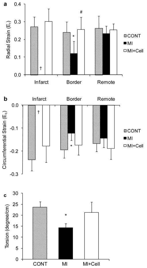
Cellular therapy for myocardial repair has been one of the most intensely investigated interventional strategies for acute myocardial infarction. Although the therapeutic potential of stem cells has been demonstrated in various studies, the underlying mechanisms for such improvements are poorly understood. In the present study, we investigated the long-term effects of stem cell therapy on both myocardial fiber organization and regional contractile function using a rat model of postinfarct remodeling. Human nonhematopoietic umbilical cord blood stem cells (nh-UCBSCs) were administered via tail vein to rats 2 days after infarct surgery. Animals were maintained without immunosuppressive therapy. In vivo and ex vivo MR imaging was performed on infarct hearts 10 months after cell transplantation. Compared to the age-matched rats exposed to the identical surgery, both global and regional cardiac functions of the nh-UCBSC-treated hearts, such as ejection fraction, ventricular strain, and torsion, were significantly improved. More importantly, the treated hearts exhibited preserved fiber orientation and water diffusivities that were similar to those in sham-operated control hearts. These data provide the first evidence that nh-UCBSC treatment may prevent/delay untoward structural remodeling in postinfarct hearts, which supports the improved LV function observed in vivo in the absence of immunosuppression, suggesting a beneficial paracrine effect occurred with the cellular therapy.
Conflict of interest statement
Authors declare no conflicts of interest.
Figures


References
-
- Amado LC, Saliaris AP, Schuleri KH, StJohn M, Xie JS, Cattaneo S, Durand DJ, Fitton T, Kuang JQ, Stewart G, Lehrke S, Baumgartner WW, Martin BJ, Heldman AW, Hare JM. Cardiac repair with intramyocardial injection of allogeneic mesenchymal stem cells after myocardial infarction. Proc Natl Acad Sci U S A. 2005;102:11474–11479. - PMC - PubMed
-
- Arts T, Reneman RS, Veenstra PC. A model of the mechanics of the left ventricle. Ann Biomed Eng. 1979;7:299–318. - PubMed
-
- Basser PJ. Inferring microstructural features and the physiological state of tissues from diffusion-weighted images. NMR Biomed. 1995;8:333–344. - PubMed
-
- Basser PJ, Pierpaoli C. Microstructural and physiological features of tissues elucidated by quantitative-diffusion-tensor MRI. J Magn Reson B. 1996;111:209–219. - PubMed
Publication types
MeSH terms
Substances
Grants and funding
LinkOut - more resources
Full Text Sources
Medical
