. 2014 Feb 20;426(4):921-35.
doi: 10.1016/j.jmb.2013.12.002. Epub 2013 Dec 9.

The glaucoma-associated olfactomedin domain of myocilin forms polymorphic fibrils that are constrained by partial unfolding and peptide sequence

Affiliations

The glaucoma-associated olfactomedin domain of myocilin forms polymorphic fibrils that are constrained by partial unfolding and peptide sequence

Shannon E Hill et al. J Mol Biol. .

Abstract

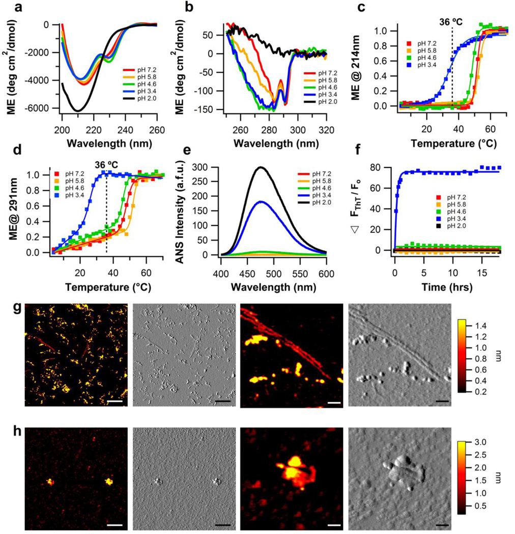
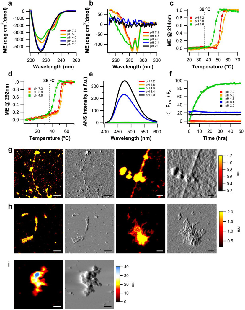
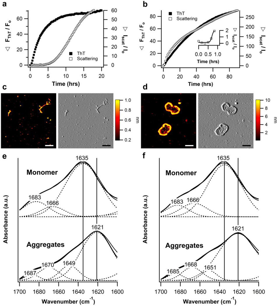
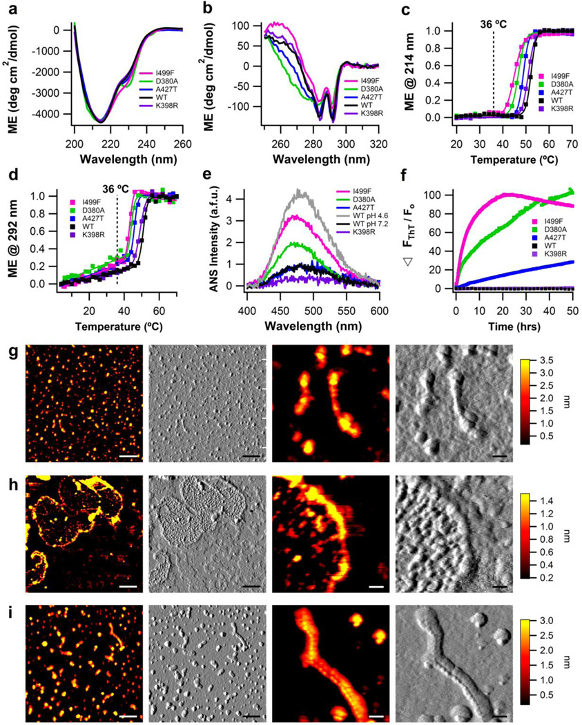
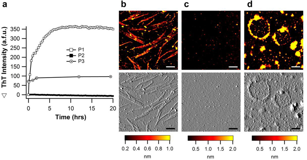
The glaucoma-associated olfactomedin domain of myocilin (myoc-OLF) is a recent addition to the growing list of disease-associated amyloidogenic proteins. Inherited, disease-causing myocilin variants aggregate intracellularly instead of being secreted to the trabecular meshwork, which is a scenario toxic to trabecular meshwork cells and leads to early onset of ocular hypertension, the major risk factor for glaucoma. Here we systematically structurally and biophysically dissected myoc-OLF to better understand its amyloidogenesis. Under mildly destabilizing conditions, wild-type myoc-OLF adopts non-native structures that readily fibrillize when incubated at a temperature just below the transition for tertiary unfolding. With buffers at physiological pH, two main endpoint fibril morphologies are observed: (a) straight fibrils common to many amyloids and (b) unique micron-length, ~300 nm or larger diameter, species that lasso oligomers, which also exhibit classical spectroscopic amyloid signatures. Three disease-causing variants investigated herein exhibit non-native tertiary structures under physiological conditions, leading to a variety of growth rates and a fibril morphologies. In particular, the well-documented D380A variant, which lacks calcium, forms large circular fibrils. Two amyloid-forming peptide stretches have been identified, one for each of the main fibril morphologies observed. Our study places myoc-OLF within the larger landscape of the amylome and provides insight into the diversity of myoc-OLF aggregation that plays a role in glaucoma pathogenesis.

Keywords: AFM; ANS; ER; FTIR; Fourier transform infrared spectroscopy; TEV; TM; ThT; amyloid; anilinonaphthalene-1-sulfonate; atomic force microscopy; circular dichroism; endoplasmic reticulum; protein misfolding; protein structure; thioflavin T; tobacco etch virus; trabecular meshwork.


Figures

Figure 1
Figure 1. Biophysical characterization of wild-type myoc-OLF as a function of pH, without NaCl
(a) Secondary structure of myoc-OLF as a function of pH measured by far-UV CD. (b) Tertiary structure of myoc-OLF as a function of pH measured by near-UV CD. (c) Thermal unfolding of secondary structure monitored by CD at 214 nm. Fit is sigmoidal. (d) Thermal unfolding of tertiary structure monitored by CD at 291 nm. Fit is linear plus sigmoidal. (e) ANS fluorescence as a function of pH. (f) ThT fluorescence as a function of pH at 36 °C monitored for 18 hours. (g) Fibrillar end point morphology for samples incubated at 36 °C in pH 3.4 buffer (h) Disordered aggregates from samples incubated at pH 2.0 buffer at 36 °C, upon deposition (see text). For (g) and (h), two left panels scale bar = 300nm and two right panels scale bar = 50nm.
Figure 2
Figure 2. Biophysical characterization of wild-type myoc-OLF as a function of pH, in the presence of 200mM NaCl
(a) Secondary structure of myoc-OLF as a function of pH measured by far-UV CD. (b) Tertiary structure of myoc-OLF as a function of pH measured by near-UV CD. (c) Thermal unfolding of secondary structure monitored by CD at 214 nm. Fit is sigmoidal. (d) Thermal unfolding of tertiary structure monitored by CD at 292 nm. Fit is linear plus sigmoidal. (e) ANS fluorescence as a function of pH. (f) ThT fluorescence as a function of pH at 36 °C monitored for 50 hours. (g) Fibrillar end point morphology for samples incubated at 36 °C in pH 4.6 buffer. (h) Deposits of curvilinear fibrils from samples incubated at 36 °C in pH 3.4 buffer. (i) Disordered aggregates for samples incubated at 36 °C in pH 2.0 buffer. For (g-i), two left panels scale bar = 300nm and two right panels scale bar = 50nm.
Figure 3
Figure 3. Fibrillization of wild-type myoc-OLF incubated at 42 °C in neutral pH buffer
(a) ThT fibril growth curves including scattering intensity kinetics for incubation at 42 °C without NaCl and (b) with 200mM NaCl. (c) Extended fibril morphology observed for samples incubated in the absence of NaCl. (d) Loop morphology observed for samples incubated in the presence of 200mM NaCl. Scale bar for both conditions (c, d) is 300nm. (e) FTIR spectral analysis for monomers and aggregates without NaCl and (f) with 200mM NaCl.
Figure 4
Figure 4. Biophysical characterization of myoc-OLF variants K398R (SNP), D380A, A427T, and I499F (disease-causing) in physiological buffers
(a) Secondary structure measured by far-UV CD. (b) Tertiary structure measured by near-UV CD. (c) Thermal unfolding of secondary structure monitored by CD at 214 nm. Fit is sigmoidal. (d) Thermal unfolding of tertiary structure monitored by CD at 292 nm. Fit is linear plus sigmoidal. For (a)-(d), wild-type myoc-OLF at neutral pH with 200 mM NaCl is overlayed for comparison. (e) ANS fluorescence as a function of pH, with overlay of wild-type myoc-OLF at pH 7.2/200 mM NaCl and 4.6/200mM NaCl. (f) ThT fluorescence at 36 °C monitored for 50 hours. (g) End-point morphologies seen for myoc-OLF(A427T) are fibrils and oligomers. (h) Deposits of myoc-OLF(D380A) appear as curvilinear and circular fibrils enclosing smaller globular aggregates. (i) Morphologies seen for myoc-OLF(I499F) are fibrils and oligomers, similar to myoc-OLF(A427T). For (g)-(i), two left panels scale bar = 300nm and two right panels scale bar = 50nm.
Figure 5
Figure 5. Fibrillization of peptides P1-P3 incubated at 36 °C in physiological buffer
(a) ThT fibril growth kinetics. (b) Straight fibril morphology observed for P1 (c) No aggregates observed for P2 (d) Closed loop morphology observed for P3. Scale bars for (b, c, d) are 300nm.

Similar articles

Cited by

References

    1. Stone EM, Fingert JH, Alward WL, Nguyen TD, Polansky JR, Sunden SL, Nishimura D, Clark AF, Nystuen A, Nichols BE, Mackey DA, Ritch R, Kalenak JW, Craven ER, Sheffield VC. Identification of a gene that causes primary open angle glaucoma. Science. 1997;275:668–670. - PubMed
    1. Gobeil S, Rodrigue M-A, Moisan S, Nguyen TD, Polansky JR, Morissette J, Raymond V. Intracellular sequestration of hetero-oligomers formed by wild-type and glaucoma-causing myocilin mutants. Invest. Ophthalmol. Vis. Sci. 2004;45:3560–3567. - PubMed
    1. Joe MK, Sohn S, Hur W, Moon Y, Choi YR, Kee C. Accumulation of mutant myocilins in ER leads to ER stress and potential cytotoxicity in human trabecular meshwork cells. Biochem. Biophys. Res. Commun. 2003;312:592–600. - PubMed
    1. Yam GH-F, Gaplovska-Kysela K, Zuber C, Roth J. Aggregated myocilin induces russell bodies and causes apoptosis: implications for the pathogenesis of myocilin-caused primary open-angle glaucoma. Am. J. Pathol. 2007;170:100–109. - PMC - PubMed
    1. Zhou Z, Vollrath D. A cellular assay distinguishes normal and mutant TIGR/myocilin protein. Hum. Mol. Genet. 1999;8:2221–2218. - PubMed

Publication types

MeSH terms