. 2014 Feb 15:223:114-22.
doi: 10.1016/j.jneumeth.2013.11.024. Epub 2013 Dec 12.

A cellular model of amyloid precursor protein processing and amyloid-β peptide production

Affiliations

A cellular model of amyloid precursor protein processing and amyloid-β peptide production

Mimi P Macias et al. J Neurosci Methods. .

Abstract

Background: A hallmark pathologic feature of Alzheimer's disease (AD) is accumulation of neuritic senile plaques in the brain parenchyma. Neurotoxic plaque cores are composed predominantly of amyloid-β (Aβ) peptides of 40 and 42 amino acids in length, formed by sequential cleavage of amyloid precursor protein (APP) by β-, and γ-secretases. There is a great interest in approaches to modulate Aβ peptide production and develop therapeutic interventions to reduce Aβ levels to halt or slow the progression of neurodegeneration.

New method: We characterized and present the BE(2)-M17 human neuroblastoma cell line as a novel in vitro model of the APP-cleavage cascade to support future (1) functional studies of molecular regulators in Aβ production, and (2) high-throughput screening assays of new pharmacotherapeutics.

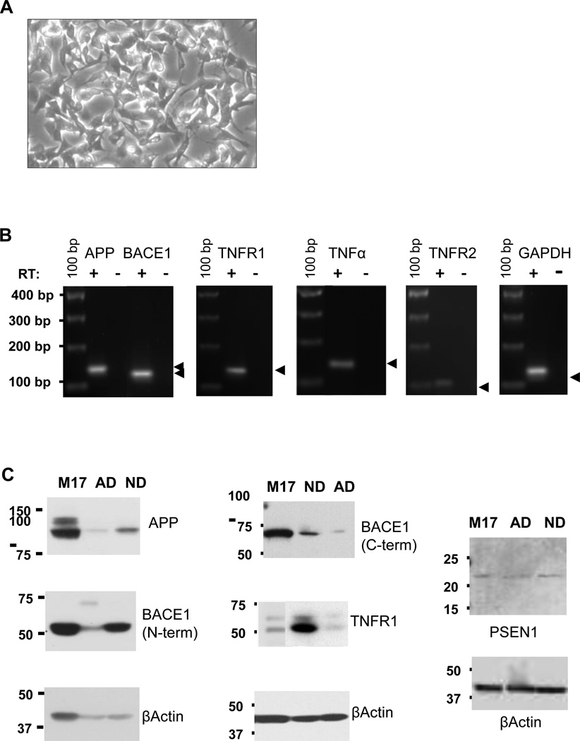
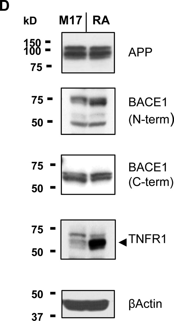
Results: In BE(2)-M17 cells, both RNA (i.e., RT-PCR, RNA sequencing) and protein analyses (i.e., Western blots, ELISA), show endogenous expression of critical components of the amyloidogenic pathway, APP-cleavage intermediates CTF83 and CTF99, and final cleavage products Aβ40 and Aβ42. We further report effects of retinoic acid-mediated differentiation on morphology and gene expression in this cell line.

Comparison with existing method(s): In contrast to primary isolates or other cell lines reported in current literature, BE(2)-M17 not only sustains baseline expression of the full contingent of APP-processing components, but also remains stably adherent during culture, facilitating experimental manipulations.

Conclusions: Our evidence supports the use of BE(2)-M17 as a novel, human, cell-based model of the APP processing pathway that offers a potential streamlined approach to dissect molecular functions of endogenous regulatory pathways, and perform mechanistic studies to identify modulators of Aβ production.

Keywords: Alzheimer's; Amyloid-beta; BE(2)-M17; In vitro; Secretase; TNF.


Conflict of interest statement

The authors declare no conflict of interest.

Figures

Fig. 1
Fig. 1. BE(2)-M17 cells
(A) Phase contrast micrograph of the neuroblastoma cell line (100× magnification). (B) Endogenous RNA gene expression in BE(2)-M17 cells by RT-PCR analysis. M17 RNA was either reverse transcribed (RT +) or for negative controls, not reverse transcribed (RT −) and the amplicons from RT-PCR visualized on 2% agarose/0.5× TBE gels: APP 141 bp; BACE1, 122 bp; TNFR1, 134 bp; TNFα, 135 bp; TNFR2, 102 bp; GAPDH, 131 bp. Amplicon positions are indicated by arrowheads to the right of each panel. 100 bp Ladder and marker sizes are shown on the left. (C) Western blots of BE(2)-M17 cell lysates (M17), Alzheimer’s (AD) and Non-Demented (ND) human brain homogenates detect expression of proteins associated with AD neuropathology. On 9% Tris-Glycine gels, 25 µg (left 3 panels) or 50 µg (center 3 panels), and on an 18% Tris-Glycine gel, 30 µg (right 2 panels) samples were electrophoretically separated and transferred to PVDF membranes for immunoblotting. Anti-βactin blots are shown as sample loading controls. Molecular weights of Precision Plus protein standards (kilodaltons) are given to the left of each panel.
Fig. 2
Fig. 2. Amyloid Precursor Protein (APP) cleavage pathway products in M17 cell lysates and conditioned media
(A) Tris-Tricine Gel Western blotting detects the presence of both β- and α-secretase-mediated APP-cleavage products, carboxy-terminal fragments CTF99 and CTF83, respectively. 100 µg of total M17 lysate was electrophoretically separated and transferred to Immobilon-PSQ PVDF membrane for immunoblot detection using an anti-APP carboxy terminal 20aa primary antibody, SIG39152. Precision Plus and Kaleidoscope molecular weight standards (left) are given for size references in kilodaltons (kD). (B) ELISA quantitation of Aβ production by BE(2)-M17 cells. Using Novex ELISA kits specific for human Aβ40 and Aβ42, levels of secreted Aβ40 and Aβ42 were measured neat in the conditioned medium of BE(2)-M17 cultures at either 48 hrs (48-M17) or 120 hrs (120-M17) post-feeding. (C) β-secretase activity in M17 cell lysates. The level of secretase enzymatic activity measured on an Abcam kit-provided substrate, is proportional to the level of fluorescent signal intensity shown on the y-axis. Total protein quantity of M17 lysate per assay is shown on the x-axis (µg).
Fig. 3
Fig. 3. Four day treatment of BE(2)-M17 cells with 10 µM retinoic acid (RA)
(A) Phase contrast microscopic images (100× magnification) of untreated M17 neuroblastoma cells (left panel) and retinoic acid-treated cells (right panel). The RA-treated cells exhibit the morphological changes, increased number of neuritic extensions, and increased length of neurite outgrowths associated with RA-mediated neuroblastoma differentiation. (B) Endpoint RT-PCR analysis of altered gene expression in untreated BE(2)-M17 (M17) and RA-treated cells (RA). Total RNAs purified from M17 or RA-treated cells were reverse transcribed (RT +) or not (RT −) for RT-PCR assays and the products separated on 2% agarose/0.5× TBE gels. Names of target genes are indicated above, and amplicon sizes indicated below, each panel. Migration of the 100 bp Ladder size standards are shown to the left. C Real-time quantitative RT-PCR (QRT-PCR) Relative Quantity Charts of 10 µM Retinoic Acid (RA)-Treated BE(2)-M17 Cells (day 4). The changes in expression of the genes-of-interest in RA-treated M17 cells, relative to untreated controls, are given in parentheses above each chart. D Western Blots of M17 lysates from untreated and retinoic acid-treated (day 4) cells. Electrophoretic separation of 100 µg of total protein of M17 or RA-treated (RA) cell lysate through 12% Tris-Glycine SDS-PAGE gels was followed by immunoblotting and ECL detection of the effects of RA-treatment on protein expression. Consistent with increased TNFR1 RNA by RT-PCR (Fig. 3B and 3C) is increased TNFR1 protein expression in RA-treated cell lysates, indicated by an arrowhead to the right of the TNFR1 panel. The anti-βactin blot is shown as a sample loading control. Positions of the Precision Plus molecular weight standards in kilodaltons (kD), are shown to the left.

References

    1. Andrau D, Dumanchin-Njock C, Aryal E, Vizzavona J, Farzan M, Boisbrun M, Fulcrand P, Hernandez J-F, Martinez J, Lefranc-Jullien S, et al. BACE1- and BACE2-expressing Human Cells. The Journal of Biological Chemistry. 2003;278(28):25859–25866. - PubMed
    1. Andres D, Keyser BM, Petrali J, Benton B, Hubbard KS, McNutt PM, Ray R. Morphological and functional differentiation in BE(2)-M17 human neuroblastoma cells by treatment with Trans-retinoic acid. BMC Neurosci. 2013;14:49. - PMC - PubMed
    1. Bien J, Jefferson T, Causevic M, Jumpertz T, Munter L, Multhaup G, Weggen S, Becker-Pauly C, Pietrzzik CU. The metalloprotease meprin β generates amino terminal-truncated amyloid β peptide species. J Biol Chem. 2012;287(40):33304–33313. - PMC - PubMed
    1. Carter DB, Dunn E, Pauley AM, McKinley DD, Fleck TJ, Ellerbrook BR, Stratman NC, Zhou X, Himes CS, Nye JS, et al. Changes in gamma-secretase activity and specificity caused by the introduction of consensus aspartyl protease active motif in Presenilin 1. Mol Neurodegener. 2008;3:6. - PMC - PubMed
    1. Chow VW, Mattson MP, Wong PC, Gleichmann M. An overview of APP processing enzymes and products. Neuromolecular Med. 2010;12(1):1–12. - PMC - PubMed

Publication types

MeSH terms