. 2014 Mar:309:124-35.
doi: 10.1016/j.heares.2013.11.009. Epub 2013 Dec 12.

Connexin 26 null mice exhibit spiral ganglion degeneration that can be blocked by BDNF gene therapy

Affiliations

Connexin 26 null mice exhibit spiral ganglion degeneration that can be blocked by BDNF gene therapy

Yohei Takada et al. Hear Res. 2014 Mar.

Abstract

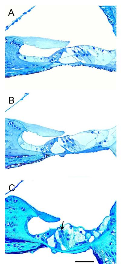
Mutations in the connexin 26 gene (GJB2) are the most common genetic cause of deafness, leading to congenital bilateral non-syndromic sensorineural hearing loss. Here we report the generation of a mouse model for a connexin 26 (Cx26) mutation, in which cre-Sox10 drives excision of the Cx26 gene from non-sensory cells flanking the auditory epithelium. We determined that these conditional knockout mice, designated Gjb2-CKO, have a severe hearing loss. Immunocytochemistry of the auditory epithelium confirmed absence of Cx26 in the non-sensory cells. Histology of the organ of Corti and the spiral ganglion neurons (SGNs) performed at ages 1, 3, or 6 months revealed that in Gjb2-CKO mice, the organ of Corti began to degenerate in the basal cochlear turn at an early stage, and the degeneration rapidly spread to the apex. In addition, the density of SGNs in Rosenthal's canal decreased rapidly along a gradient from the base of the cochlea to the apex, where some SGNs survived until at least 6 months of age. Surviving neurons often clustered together and formed clumps of cells in the canal. We then assessed the influence of brain derived neurotrophic factor (BDNF) gene therapy on the SGNs of Gjb2-CKO mice by inoculating Adenovirus with the BDNF gene insert (Ad.BDNF) into the base of the cochlea via the scala tympani or scala media. We determined that over-expression of BDNF beginning around 1 month of age resulted in a significant rescue of neurons in Rosenthal's canal of the cochlear basal turn but not in the middle or apical portions. This data may be used to design therapies for enhancing the SGN physiological status in all GJB2 patients and especially in a sub-group of GJB2 patients where the hearing loss progresses due to ongoing degeneration of the auditory nerve, thereby improving the outcome of cochlear implant therapy in these ears.


Figures

Figure 1
Figure 1
Epi-fluorescence of whole mounts (middle turn) showing C×26 immunoreactivity (red) and phalloidin (green) (A-C) and ABR thresholds recorded at 1 month of age in wild type and mutant mice (D). (A-B) In wild type auditory epithelium C×26 immunoreactivity appears as a conspicuous dotted line surrounding apical surfaces of non-sensory cells. The brightest staining was found in the inner sulcus cell region. Panel B is a high-magnification images of the inset in A. (C) In a mutant cochlea, C×26 immunoreactivity is absent in the inner sulcus and other regions. Hair cells are missing and supporting cells are present in one area (on left) but no in other areas, where they are replaced by a flat epithelium. Scale bars indicate 50 μm. (D) ABR thresholds at 12, 16, and 24 kHz in wild type and mutant mice measured at 1 month of age. Auditory thresholds were significantly higher in Gjb2-CKO mice (p<0.001) than in wild type mice at 12 and 16 and 24 kHz. Sample size for Gjb2-CKO mice: n=8 at 12 & 16 kHz, n=5 at 24 kHz (thresholds for 3 mice could not be detected at the maximum stimulation level of 110 dB). Sample size for wild type mice: n=4 at all three frequencies. Error bars indicate the standard deviation of the mean.
Figure 2
Figure 2
A panel of photomicrographs of cross sections through the organ of Corti of wild type mice at 6 months of age. (A-B) The organ of Corti region in the apex (A) and middle (B) contain the normal cellular components including hair cells and supporting cells and appears normally organized. (C) In the base area the inner hair cell appears normal but partial outer hair cells degeneration is observed (arrow). Scale bar indicates 50 μm.
Figure 3
Figure 3
Cross sections through the organ of Corti of mutant mice from ears obtained at ages 1 (A-C), 3 (D-F) or 6 months (G-I). (A-B) In the apex (A) and middle (B), the organ of Corti region displays partial degeneration of hair cells and the tunnel of Corti appears poorly formed. Supporting cells remain in a differentiated state. (C) The organ of Corti region in the base is completely replaced by a flat epithelium. (D) The organ of Corti region in the apex is similar to 1 month ears (A), but the organ of Corti region in the middle (E) and base (F) is replaced by flat epithelium. (G-I) At 6 months of age, the organ of Corti region in all turns is lacking sensory cells and differentiated supporting cells. The tissue appears as a flat epithelium throughout the cochlea. Scale bar indicates 50 μm.
Figure 4
Figure 4
Photomicrographs of cross sections through Rosenthal’s canal of wild type mice from ears obtained at ages 1 (A-C), 3 (D- F) or 6 months (G-I). (A-F) Rosenthal’s canal is occupied by soma of SGNs in all cochlear turns. Neurons appear normal but occasional clustering is observed. (G-I) The SGN density appears reduced in all cochlear turns. In the apex (G) somata of several SGNs are clustered together. Scale bar indicates 50 μm.
Figure 5
Figure 5
Cross sections through Rosenthal’s canal of mutant mice (A-I) from ears obtained at ages 1 (A-C), 3 (D-F) or 6 months (G-I). (A-B) Rosenthal’s canal is fully occupied by soma of SGNs in the apex and middle cochlear turns. (C) The SGN density appears reduced in the base. (D) The SGN density in apex is similar to 1 month ears (A). (E-F) The SGN density is reduced markedly from base (F) to middle (E). (G-I) At 6 months, the SGN density is reduced in all cochlear turns. Soma of surviving SGNs are often clustered together to form isolated islands in Rosenthal’s canal (G). Scale bar indicates 50 μm.
Figure 6
Figure 6
Comparison of SGN density between wild type and mutant mice. There is a significant difference in density of SGNs between the ages of 1 month and 6 months. In the mutant mice, these data show that SGN degeneration progressed from base to apex gradually. Data in wild type ears show that SGN degeneration starts to manifest at 6 months in all turns, caused by the age-related hearing loss mutation in C57BL/6 mice. Error bars indicate 1 standard error of the mean.
Figure 7
Figure 7
(A-B): Sections of representative Rosenthal’s canals of 2 month old mice, in ears treated with Ad.BDNF into the perilymph (B) or contralateral ears (A). (A) The SGNs in the base of contralateral ears were very sparse as most cells have degenerated by this stage. Surviving cells appear pyknotic. (B) SGNs in ears treated with Ad.BDNF appeared healthy and occupied most of the space in Rosenthal’s canal. Connective tissue could be observed in the scala tympani (arrowhead). (C) The density of SGNs in Rosenthal’s canal after Ad.BDNF was inoculated into the scala tympani, compared to untreated (contralateral) ears. The density of SGNs in the Ad.BDNF-treated-ears was significantly different from contralateral ears only in the basal profile of Rosenthal’s canal (*, p< 0.05). Bars are standard error of the mean and statistical comparison was done by t-test for paired samples (N=5). Scale bar indicates 50 μm.
Figure 8
Figure 8
(A-B): Sections of representative profiles of Rosenthal’s canal of 2 month old mice, in ears treated with Ad.BDNF into the endolymph (B) or contralateral ears (A). (A) The SGNs in the basal turn of contralateral ears were very sparse and pyknotic. (B) SGNs in ears treated with Ad.BDNF into the endolymph appeared large and almost fully occupied the canal. Connective tissue could be observed in the scala tympani (arrowhead). (C) Quantitative analyses of the density of SGNs in Rosenthal’s canal after Ad.BDNF inoculation into the scala media compared to untreated (contralateral) ears. The density of SGNs in the Ad.BDNF-treated ears was significantly greater than contralateral ears only in the basal profile of Rosenthal’s canal (*, p< 0.05). Bars are standard error of the mean and statistical comparison was done by t-test for paired samples (N=9). Scale bar indicates 50 μm.

Similar articles

Cited by

References

    1. Aarnisalo AA, Pirvola U, Liang XQ, Miller J, Ylikoski J. Apoptosis in auditory brainstem neurons after a severe noise trauma of the organ of Corti: intracochlear GDNF treatment reduces the number of apoptotic cells. ORL. J. Otorhinolaryngol. Relat. Spec. 2000;62:330–4. - PubMed
    1. Abrashkin KA, Izumikawa M, Miyazawa T, Wang CH, Crumling MA, Swiderski DL, Beyer LA, Gong TW, Raphael Y. The fate of outer hair cells after acoustic or ototoxic insults. Hear. Res. 2006;218:20–9. - PubMed
    1. Agterberg MJ, Versnel H, van Dijk LM, de Groot JC, Klis SF. Enhanced survival of spiral ganglion cells after cessation of treatment with brain-derived neurotrophic factor in deafened guinea pigs. J Assoc Res Otolaryngol. 2009;10:355–67. - PMC - PubMed
    1. Agterberg MJ, Versnel H, de Groot JC, Smoorenburg GF, Albers FW, Klis SF. Morphological changes in spiral ganglion cells after intracochlear application of brain-derived neurotrophic factor in deafened guinea pigs. Hear. Res. 2008;244:25–34. - PubMed
    1. Anselmi F, Hernandez VH, Crispino G, Seydel A, Ortolano S, Roper SD, Kessaris N, Richardson W, Rickheit G, Filippov MA, Monyer H, Mammano F. ATP release through connexin hemichannels and gap junction transfer of second messengers propagate Ca2+ signals across the inner ear. Proc. Natl. Acad. Sci. U. S. A. 2008;105:18770–5. - PMC - PubMed

Publication types

MeSH terms