. 2014 Feb 15;321(2):109-22.
doi: 10.1016/j.yexcr.2013.11.023. Epub 2013 Dec 10.

The regulation of RhoA at focal adhesions by StarD13 is important for astrocytoma cell motility

Affiliations

The regulation of RhoA at focal adhesions by StarD13 is important for astrocytoma cell motility

Bassem D Khalil et al. Exp Cell Res. .

Abstract

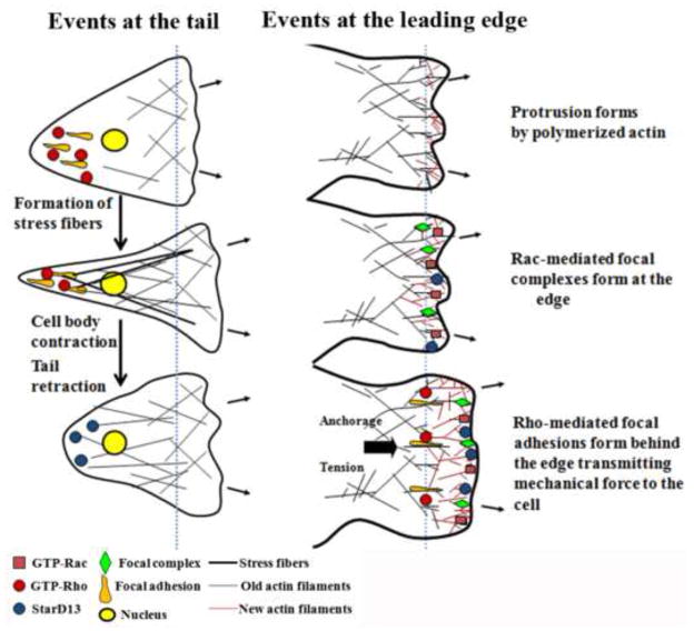
Malignant astrocytomas are highly invasive into adjacent and distant regions of the normal brain. Rho GTPases are small monomeric G proteins that play important roles in cytoskeleton rearrangement, cell motility, and tumor invasion. In the present study, we show that the knock down of StarD13, a GTPase activating protein (GAP) for RhoA and Cdc42, inhibits astrocytoma cell migration through modulating focal adhesion dynamics and cell adhesion. This effect is mediated by the resulting constitutive activation of RhoA and the subsequent indirect inhibition of Rac. Using Total Internal Reflection Fluorescence (TIRF)-based Förster Resonance Energy Transfer (FRET), we show that RhoA activity localizes with focal adhesions at the basal surface of astrocytoma cells. Moreover, the knock down of StarD13 inhibits the cycling of RhoA activation at the rear edge of cells, which makes them defective in retracting their tail. This study highlights the importance of the regulation of RhoA activity in focal adhesions of astrocytoma cells and establishes StarD13 as a GAP playing a major role in this process.

Keywords: Astrocytoma; Cell motility; Rac; RhoA; StarD13.


Figures

Figure 1
Figure 1. StarD13 is needed for cell motility
Cells (T98G) were transfected with luciferase control siRNA or with StarD13 siRNA (2 oligos) for 72 hours. A) The cells were lysed and immunoblotted by western blot analysis for StarD13 (upper gel) or for actin (lower gel) for loading control. B) Cells transfected with luciferase siRNA, StarD13 siRNA, or StarD13 siRNA and GFP-StarD13 were grown in a monolayer then wounded and left to recover the wound then imaged at the same frame after 24 hours (lower micrographs). Scale bar is 50 μm. C) Quantitation for B). Wound widths were measured at 11 different points for each wound, and the average rate of wound closure for the cells was calculated in μm/hr. Data are the mean −/+ SEM from 3 wound closure movies. ** indicates that the value is significant with p<0.01. D) The net paths of projected 120 frames from 2 hour long time lapse movies of cells (SF268) transfected with luciferase control siRNA or with StarD13 siRNA undergoing random motility in serum (different colors represent different cells) E) Quantitation of the cell speed from D) expressed in μm/min or the net paths shown expressed in μm. Data are the mean −/+ SEM from 15 cells. *** indicates that the values are significant with p<0.001.
Figure 2
Figure 2. Proper regulation by StarD13 of Rho GTPases is needed for adhesion dynamics and tail detachment
A) Representative micrographs of cells (T98G) transfected with luciferase control, StarD13 siRNA or StarD13 siRNA and GFP-StarD13 (upper panel) or RhoA and Rac siRNA (lower panel) that were fixed and immunostained with paxillin. B) Quantitation of focal adhesion size in cells transfected with luciferase control or StarD13 siRNA. Data are the mean +/− SEM from 3 different experiments. C) Representative fluorescent micrographs taken from a time lapse of cells were transfected with luciferase or StarD13 siRNA and then transfected with GFP-vinculin and imaged while undergoing random motility in serum for 1 hour. D) Quantitation of C) showing percentage of focal adhesions persisting more than 20 minutes (Control n=32 and StarD13 siRNA n=23). E) Representative phase contrast micrographs taken from a time lapse of cells were transfected with luciferase or StarD13 siRNA and imaged while undergoing random motility in serum for 2 hours. F) Representative micrographs of cells fixed and stained with crystal violet to detect adhesion (as described in methods). Scale bar is 50 μm. G) Graph shows quantitation of F) where crystal violet was solubilized and the absorption of the plates was read at 550 nm using an ELISA plate reader. Data is measured in arbitrary units and normalized to the luciferase control. Data are the mean −/+ SEM from 3 experiments (15 cells/condition/experiment). ** indicates that the value is significant with p=0.007.
Figure 3
Figure 3. Effect of StarD13-RhoA on Rac activation which is needed for cell motility
A) T98G cells were transfected with luciferase, StarD13, RhoA, or StarD13 and RhoA siRNA for 72 hours. The cells were then lysed and incubated with GST-RBD (Rhotekin binding domain), or with GST-CRIB (Cdc42 and Rac interactive binding domain) to pull down active RhoA or Rac respectively. The samples were then blotted with RhoA or Rac antibody. The lower gel in each panel is a western blot for the total cell lysates for loading control. B) Cells (T98G) were transfected with luciferase control siRNA or with Rac siRNA for 72 hours. The cells were lysed and immunoblotted by western blot analysis for Rac (upper gel) or for actin (lower gel) for loading control. C) The luciferase siRNA-transfected and Rac siRNA-transfected cells were grown in a monolayer then wounded and left to recover the wound then imaged at the same frame after 24 hours (lower micrographs). Scale bar is 50 μm. D) Quantitation for C). Wound widths were measured at 11 different points for each wound, and the average rate of wound closure for the cells was calculated in μm/hr. Data are the mean −/+ SEM from 3 wound closure movies. ** indicates that the value is significant with p=0.001. E) The net paths of projected 120 frames from 2 hour long time lapse movies of cells (SF268) transfected with luciferase control siRNA or with Rac siRNA undergoing random motility in serum (different colors represent different cells) F) Quantitation of the cell speed from F) expressed in μm/min or the net paths shown expressed in μm. Data are the mean −/+ SEM from 15 cells. *** indicates that the values are significant with p<0.001.
Figure 4
Figure 4. Constitutively active RhoA inhibits cell motility
A) Cells (T98G) were transfected with GFP vector or dominant active RhoA construct (RhoA DA). Cells were grown in a monolayer then wounded and left to recover the wound then imaged at the same frame after 24 hours (lower micrographs). B) Quantitation for A). Wound widths were measured at 11 different points for each wound, and the average rate of wound closure was calculated in μm/hr. Data are the mean −/+ SEM from 3 wound closure movies. * indicates that the value is significant with p=0.01. C) Cells (SF268) were transfected with GFP vector or with GFP-RhoA-DA. The images show net paths of projected 120 frames from 2 hour long time lapse movies of green cells undergoing random motility in serum (different colors represent different cells). D) Quantitation of the cell speed from D) expressed in μm/min or the net paths shown expressed in μm. Data are the mean −/+ SEM from 15 cells. *** indicates that the values are significant with p<0.001.
Figure 5
Figure 5. StarD13 regulation of focal adhesions is needed for cell motility
A) Cells were transfected with luciferase control, StarD13 and dominant active Rac, StarD13 and RhoA siRNA, or StarD13, Rac, and RhoA siRNA, lysed and western blots of total lysates blotted with StarD13, RhoA, Rac or actin antibodies. B) Same cells as in A) transfected and grown in a monolayer then wounded and left to recover the wound then imaged at the same frame after 24 hours (lower micrographs). C) Quantitation for B). Wound widths were measured at 11 different points for each wound, and the average rate of wound closure for the cells was calculated in μm/hr. Data are the mean −/+ SEM from 3 wound closure movies. ** indicates that the values are significant with p<0.01. D) Representative micrographs of cells (T98G) transfected with luciferase control, StarD13 and Rac siRNA, StarD13 and RhoA siRNA, or StarD13, Rac, and RhoA siRNA that were fixed and immunostained with paxillin. Scale bar is 10 μm.
Figure 6
Figure 6. StarD13 knockdown inhibits cycling of RhoA activity and RhoA actication co-localizes with focal adhesions
A) Montage of representative SF268 cells (15 cells/condition/experiment of 3 experiments) transfected with luciferase or StarD13 siRNA and with the RhoA FRET biosensor undergoing motility in serum with the direction of migration indicated by an arrow (frames are 4 min apart). The lower panels for each cell types are a closeup of the area enclosed by a white box in the upper panels. B) The graph is a quantitation of the FRET signal in the area indicated by the shadowed area in the lower panels representing the cell tail. C) SF268 cells were transfected with 500ng of the RhoA biosensor plasmid and 500ng of the mCherry-Paxillin plasmid. The paxillin signal and the FRET signal were collected on a TIRF station.
Figure 7
Figure 7
Model of StarD13 regulation of RhoA activation at the tail and at the leading edge of astrocytoma cells undergoing cell motility.

Similar articles

Cited by

References

    1. Louis DN, Ohgaki H, Wiestler OD, Cavenee WK. WHO Classification of Tumours of the Central Nervous System. World Health Organization; 2007. - PMC - PubMed
    1. CBTRUS. CBTRUS Statistical Report: Primary Brain and Central Nervous System Tumors Diagnosed in the United States in 2004–2007. Central Brain Tumor Registry of the United States; Hinsdale, IL: 2011.
    1. Wen PY, Kesari S. Malignant gliomas in adults. N Engl J Med. 2008;359:492–507. - PubMed
    1. Nakada M, Nakada S, Demuth T, Tran NL, Hoelzinger DB, Berens ME. Molecular targets of glioma invasion. Cell Mol Life Sci. 2007;64:458–478. - PMC - PubMed
    1. Giese A, Bjerkvig R, Berens ME, Westphal M. Cost of migration: invasion of malignant gliomas and implications for treatment. J Clin Oncol. 2003;21:1624–1636. - PubMed

Publication types

MeSH terms