

Dexamethasone exacerbates cerebral edema and brain injury following lithium-pilocarpine induced status epilepticus
- PMID: 24333865
- PMCID: PMC3905166
- DOI: 10.1016/j.nbd.2013.12.001
Dexamethasone exacerbates cerebral edema and brain injury following lithium-pilocarpine induced status epilepticus
Abstract
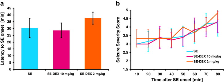
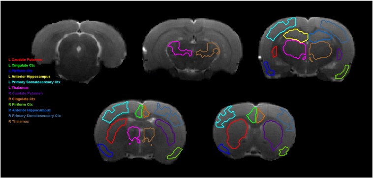
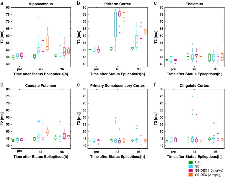
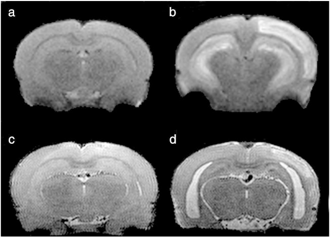
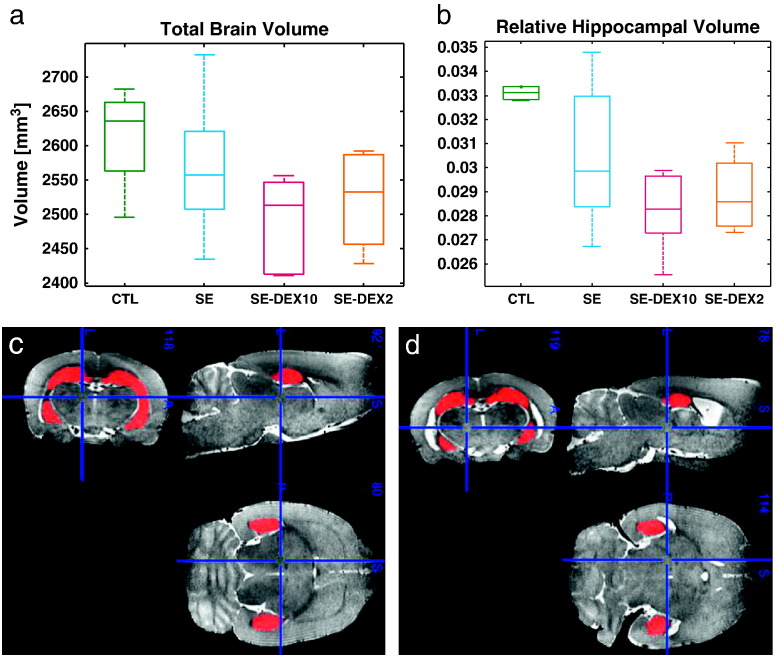
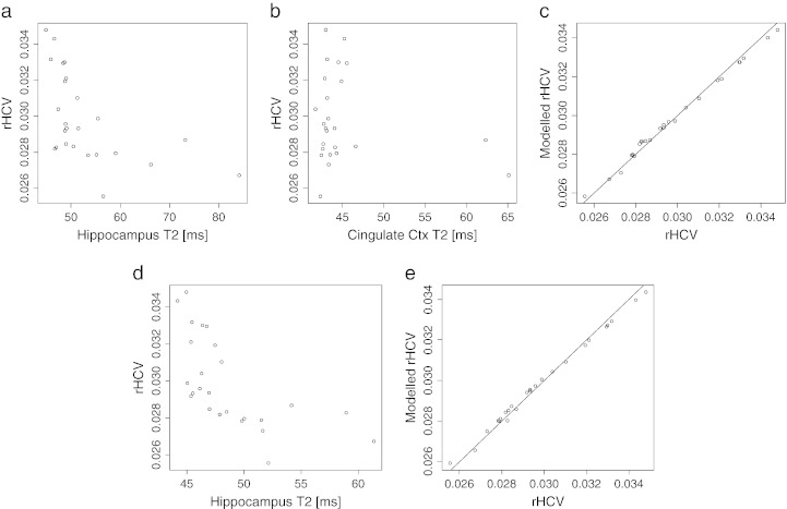
Anti-inflammatory therapies are the current most plausible drug candidates for anti-epileptogenesis and neuroprotection following prolonged seizures. Given that vasogenic edema is widely considered to be detrimental for outcome following status epilepticus, the anti-inflammatory agent dexamethasone is sometimes used in clinic for alleviating cerebral edema. In this study we perform longitudinal magnetic resonance imaging in order to assess the contribution of dexamethasone on cerebral edema and subsequent neuroprotection following status epilepticus. Lithium-pilocarpine was used to induce status epilepticus in rats. Following status epilepticus, rats were either post-treated with saline or with dexamethasone sodium phosphate (10mg/kg or 2mg/kg). Brain edema was assessed by means of magnetic resonance imaging (T2 relaxometry) and hippocampal volumetry was used as a marker of neuronal injury. T2 relaxometry was performed prior to, 48 h and 96 h following status epilepticus. Volume measurements were performed between 18 and 21 days after status epilepticus. Unexpectedly, cerebral edema was worse in rats that were treated with dexamethasone compared to controls. Furthermore, dexamethasone treated rats had lower hippocampal volumes compared to controls 3 weeks after the initial insult. The T2 measurements at 2 days and 4 days in the hippocampus correlated with hippocampal volumes at 3 weeks. Finally, the mortality rate in the first week following status epilepticus increased from 14% in untreated rats to 33% and 46% in rats treated with 2mg/kg and 10mg/kg dexamethasone respectively. These findings suggest that dexamethasone can exacerbate the acute cerebral edema and brain injury associated with status epilepticus.
Keywords: BBB; Biomarker; COX-2; CSE; Corticosteroids; DEX; Epilepsy; FOV; Inflammation; IκB; MRI; NSAIDs; ROIs; SE; T(2); TE; TEeff; TR; blood–brain barrier; convulsive status epilepticus; cyclooxygenase-2; dexamethasone; echo time; echo-train length; effective echo time; etl; fast spin-echo; field of view; fse; inhibitor of kappa-B; magnetic resonance imaging; non-steroidal anti-inflammatory drugs; rHCV; regions of interest; relative hippocampal volume; repetition time; status epilepticus; transverse magnetisation relaxation time constant.
Copyright © 2013. Published by Elsevier Inc.
Figures
Similar articles
-
Prevention of status epilepticus-induced brain edema and neuronal cell loss by repeated treatment with high-dose levetiracetam.Brain Res. 2015 May 22;1608:225-34. doi: 10.1016/j.brainres.2015.03.005. Epub 2015 Mar 11. Brain Res. 2015. PMID: 25770058
-
Astroglial loss and edema formation in the rat piriform cortex and hippocampus following pilocarpine-induced status epilepticus.J Comp Neurol. 2010 Nov 15;518(22):4612-28. doi: 10.1002/cne.22482. J Comp Neurol. 2010. PMID: 20886625
-
4,4'-Diisothiocyanatostilbene-2,2'-disulfonic acid attenuates spontaneous recurrent seizures and vasogenic edema following lithium-pilocarpine induced status epilepticus.Neurosci Lett. 2017 Jul 13;653:51-57. doi: 10.1016/j.neulet.2017.05.015. Epub 2017 May 10. Neurosci Lett. 2017. PMID: 28501694
-
What are the effects of prolonged seizures in the brain?Epileptic Disord. 2014 Oct;16 Spec No 1(Spec No 1):S6-11. doi: 10.1684/epd.2014.0689. Epileptic Disord. 2014. PMID: 25323416 Free PMC article. Review.
-
The applications of the pilocarpine animal model of status epilepticus: 40 years of progress (1983-2023).Behav Brain Res. 2023 Aug 24;452:114551. doi: 10.1016/j.bbr.2023.114551. Epub 2023 Jun 21. Behav Brain Res. 2023. PMID: 37348654 Review.
Cited by
-
Neuroinflammation as a Therapeutic Target for Mitigating the Long-Term Consequences of Acute Organophosphate Intoxication.Front Pharmacol. 2021 May 12;12:674325. doi: 10.3389/fphar.2021.674325. eCollection 2021. Front Pharmacol. 2021. PMID: 34054549 Free PMC article. Review.
-
A Commentary on Electrographic Seizure Management and Clinical Outcomes in Critically Ill Children.Children (Basel). 2023 Jan 31;10(2):258. doi: 10.3390/children10020258. Children (Basel). 2023. PMID: 36832387 Free PMC article.
-
Hippocampal glucocorticoid receptors modulate status epilepticus severity.Neurobiol Dis. 2023 Mar;178:106014. doi: 10.1016/j.nbd.2023.106014. Epub 2023 Jan 23. Neurobiol Dis. 2023. PMID: 36702319 Free PMC article.
-
Disease progression and brain atrophy in NMDAR encephalitis: Associated factor & clinical implication.Ann Clin Transl Neurol. 2022 Jul;9(7):912-924. doi: 10.1002/acn3.51604. Epub 2022 Jun 17. Ann Clin Transl Neurol. 2022. PMID: 35715951 Free PMC article.
-
Degradation of dexamethasone by acclimated strain of Pseudomonas Alcaligenes.Int J Clin Exp Med. 2015 Jul 15;8(7):10971-8. eCollection 2015. Int J Clin Exp Med. 2015. PMID: 26379892 Free PMC article.
References
-
- Al-Shorbagy M.Y. Diverse effects of variant doses of dexamethasone in lithium-pilocarpine induced seizures in rats. Can. J. Physiol. Pharmacol. 2012;90:13–21. - PubMed
-
- Altman D. Effects of dexamethasone in hypoxic–ischemic brain injury in the neonatal rat. Neonatology. 1984;46:149–156. - PubMed
-
- Auphan N. Immunosuppression by glucocorticoids—inhibition of NF-kappa-B activity through induction of I-kappa-B synthesis. Science. 1995;270:286–290. - PubMed
-
- Baik E.J. Cyclooxygenase-2 selective inhibitors aggravate kainic acid induced seizure and neuronal cell death in the hippocampus. Brain Res. 1999;843:118–129. - PubMed
Publication types
MeSH terms
Substances
Grants and funding
LinkOut - more resources
Full Text Sources
Other Literature Sources
Research Materials
