. 2014 Feb 28;114(5):833-44.
doi: 10.1161/CIRCRESAHA.114.302766. Epub 2013 Dec 13.

β-arrestin1-biased β1-adrenergic receptor signaling regulates microRNA processing

Affiliations

β-arrestin1-biased β1-adrenergic receptor signaling regulates microRNA processing

Il-Man Kim et al. Circ Res. .

Abstract

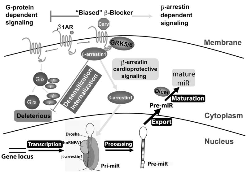
Rationale: MicroRNAs (miRs) are small, noncoding RNAs that function to post-transcriptionally regulate gene expression. First transcribed as long primary miR transcripts (pri-miRs), they are enzymatically processed in the nucleus by Drosha into hairpin intermediate miRs (pre-miRs) and further processed in the cytoplasm by Dicer into mature miRs where they regulate cellular processes after activation by a variety of signals such as those stimulated by β-adrenergic receptors (βARs). Initially discovered to desensitize βAR signaling, β-arrestins are now appreciated to transduce multiple effector pathways independent of G-protein-mediated second messenger accumulation, a concept known as biased signaling. We previously showed that the β-arrestin-biased βAR agonist, carvedilol, activates cellular pathways in the heart.

Objective: Here, we tested whether carvedilol could activate β-arrestin-mediated miR maturation, thereby providing a novel potential mechanism for its cardioprotective effects.

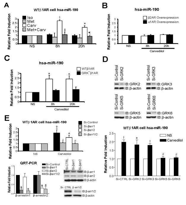
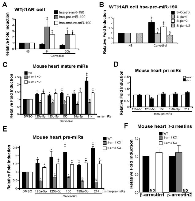
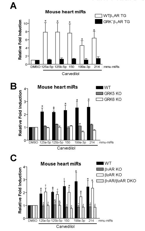
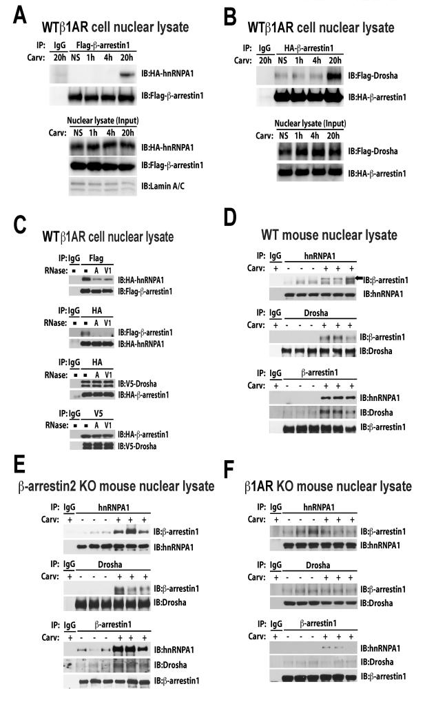
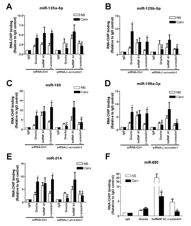
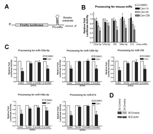
Methods and results: In human cells and mouse hearts, carvedilol upregulates a subset of mature and pre-miRs, but not their pri-miRs, in β1AR-, G-protein-coupled receptor kinase 5/6-, and β-arrestin1-dependent manner. Mechanistically, β-arrestin1 regulates miR processing by forming a nuclear complex with hnRNPA1 and Drosha on pri-miRs.

Conclusions: Our findings indicate a novel function for β1AR-mediated β-arrestin1 signaling activated by carvedilol in miR biogenesis, which may be linked, in part, to its mechanism for cell survival.

Keywords: carvedilol; heart diseases.


Figures

Figure 1
Figure 1. Carvedilol stimulation induces upregulation of human miR-190, which is dependent on β1AR, GRK5/6 and β-arrestin1
A, HEK293 cells stably expressing WTβ1ARs (WTβ1AR cells) were treated with 1μM of isoproterenol (Iso), metoprolol (Met) or carvedilol (Carv) for 8h or 20h. Expression of mature human (hsa: homo sapiens)-miR-190 was detected using the TaqMan miR assay. Among the 3 βAR ligands tested, only the β-arrestin-biased ligand Carv activated expression of hsa-miR-190. The pretreatment of 10μM Met for 4h blocked Carv-mediated hsa-miR-190 activation. B, HEK293 cells overexpressing β2ARs or α1ARs were treated with 1μM Carv and the expression of hsa-miR-190 was measured as described above. Carv did not induce miR-190 expression in β2AR- or α1AR-overexpressing cells. C, HEK293 cells stably expressing WTβ1AR or GRKβ1AR were treated with 1μM Carv. WTβ1AR induced an increase in hsa-miR-190 expression following Carv treatment, while GRKβ1AR lacked this effect. D, WTβ1AR cells were transfected with either scrambled siRNA (Si-Control or Si-CTRL) or siRNAs targeting GRKs. Hsa-miR-190 activation was abolished in cells transfected with siRNAs targeting GRK5 or GRK6. E, WTβ1AR cells were transfected with either Si-Control or siRNAs targeting β-arrestin1/2 (Si-βarr1/2), β-arrestin1 (Si-βarr1) or β-arrestin2 (Si-βarr2). Carv-mediated hsa-miR-190 activation was diminished in cells transfected with Si-β-arrestin1 or Si-β-arrestin1/2. Knockdown of β-arrestins was confirmed by both QRT-PCR and IB. NS: no stimulation with Carv (vehicle, 0.1% [v/v] DMSO). IB: immunoblotting. Data are shown as mean ± SEM for n=4 independently obtained biological samples (A, C and D) and n=5 independently obtained biological samples (B and E). *, P < 0.05 vs. NS, Iso, Met, GRK β1AR or Si-Control; †, P < 0.01 vs. NS, Iso or Met; ‡, P < 0.001 vs. NS; #, P < 0.05 vs. Si-Control; §, P < 0.01 vs. Carv; ¶, P < 0.001 vs. Carv.
Figure 2
Figure 2. β-arrestin1 is required for Carv-mediated miR activation, which occurs at a post-transcriptional step
A, WTβ1AR cells were treated with 1μM Carv for 8h or 20h. Expression of primary (pri), premature (pre), or mature hsa-miR-190 was detected using TaqMan miR assay for mature and pri-miRs and using Power SYBR Green PCR assay with pre-miR primers. Carv stimulation did not activate hsa-pri-miR-190 expression but resulted in mature or pre-hsa-miR-190 activation. B, WTβ1AR cells were transfected with siRNAs as described in Figure 1E. Carv-mediated hsa-pre-miR-190 activation was diminished in cells transfected with siRNAs targeting β-arrestin1/2 or β-arrestin1. C and E, WT, β-arrestin1 knockout (KO) or β-arrestin2 KO mice were infused with DMSO (vehicle control) or Carv (19mg/Kg/day) for 7 days by using micro-osmotic pumps. QRT-PCR experiments were performed on RNAs from mouse hearts. Five mature (C) or pre- (E) miRs were elevated upon Carv stimulation in both WT and β-arrestin2 KO mice. However, this induction was completely abolished in β-arrestin1 KO mice, indicating an essential role of β-arrestin1 in the synthesis of pre-miRs. D, WT mice were infused with DMSO or Carv as above. QRT-PCR experiments were performed in mouse hearts using Taqman pri-miR assays. Expression of pri-miRs was not changed significantly upon Carv stimulation. F, QRT-PCR analysis was performed in mouse hearts using Taqman gene expression assays for β-arrestins. NS: no stimulation. ND: not detected. Data are shown as mean ± SEM for n=7 independently obtained biological samples (A), n=5 independently obtained biological samples (B) and n=8 independent mice per group (C, D, E and F). *, P < 0.05 vs. NS, hsa-pri-miR-190 or DMSO; †, P < 0.01 vs. NS or DMSO; ‡, P < 0.001 vs. NS or DMSO; #, P < 0.05 vs. WT or β-arrestin2 KO; §, P < 0.01 vs. Si-Control, WT or β-arrestin2 KO; ¶, P < 0.01 vs. WT or β-arrestin2 KO. Notably, the levels of mature miR-214 (C) and pre-miR-214 (E) are reduced in β-arrestin2 KO compared to WT (P < 0.001) and the level of pre-miR-150 (E) is reduced β-arrestin2 KO compared to WT (P < 0.05).
Figure 3
Figure 3. Carv-mediated in vivo miR activation requires GRK5/6 phosphorylation of β1AR
A, Cardiac specific transgenic (TG) mice expressing WTβ1AR or GRK β1AR were treated with Carv as shown in Figure 2. WTβ1AR induced an increase in mature miR expression following Carv treatment, while GRK β1AR lacked this effect. B-C, WT and various KO mice were treated with Carv as aforementioned. Carv-mediated miR activation, which is seen in WT and β2AR KO mice, was abolished in GRK5 KO, GRK6 KO, β1AR KO and β1AR/β2AR double KO (DKO) mice. Data are shown as mean ± SEM for n=6 independent mice per group. *, P < 0.05 vs. DMSO or GRK β1AR TG; †, P < 0.01 vs. DMSO ; ‡, P < 0.001 vs. DMSO; #, P < 0.05 vs. WT or β2AR KO; §, P < 0.01 vs. WT or β2AR KO.
Figure 4
Figure 4. Carv induces the RNA-dependent nuclear interaction of β-arrestin1 with hnRNPA1, an important regulator of RNA helicase-independent miR processing by Drosha
A-B, WTβ1AR cells were transfected with Flag-β-arrestin1 and HA-hnRNPA1 (A) or HA-β-arrestin1 and Flag-Drosha constructs (B). After Carv treatment, nuclear extracts (NEs) were prepared and subjected to immunoprecipitation (IP) with anti-Flag, anti-HA, or non-specific IgG (control). NEs were immunobloted with lamin A/C antibody for nuclear marker. Interaction of hnRNPA1 or Drosha with β-arrestin1 was examined by immunoblotting (IB) with anti-HA and anti-Flag. C, RNA dependence of interaction of β-arrestin1 with hnRNPA1 and Drosha. WTβ1AR cells transfected with tagged plasmids were serum-starved for 4h and stimulated with Carv for 20h. NEs were treated with RNase A (single-stranded RNA nuclease) or RNase V1 (double-stranded RNA nuclease) prior to IP. Immunoprecipitates were subjected to IB. D-F, WT (D), β-arrestin2 KO (E) and β1AR KO (F) mice were infused with Carv or vehicle control and then NEs were prepared from three independent mice per group. Endogenous interaction was confirmed using indicated antibodies. NS: no stimulation.
Figure 5
Figure 5. β-arrestin1 associates with pri-β1-miRs in a Carv-dependent manner
WTβ1AR cells were transfected with pCMV-β1-miRs (A-E) or pCMV-miR-690 [control miR] (F), tagged plasmids (HA-β-arrestin1, Flag-hnRNPA1 or V5-Drosha) and control siRNA (siRNA-Ctrl) or siRNA-β-arrestin1 (A-E, as described in Figure 1E), followed by Carv treatment (20h). RNA-ChIP was performed with HA, V5, hnRNPA1 antibody or non-specific IgG (control), followed by PCR amplification with β1-miR primers (A-E) or miR-690 primers (F). Data are shown as mean ± SEM for n=4 independently obtained biological samples. *, P < 0.05 vs. NS; #, P < 0.05 vs. Carv of siRNA-Ctrl.
Figure 6
Figure 6. Carv facilitates Drosha-mediated miR processing by β-arrestin1
A, Pri-miRs of five mouse β-arrestin1-regulated miRs were cloned into 3’UTR of luciferase construct. B-D, In vivo pri-miR processing assay measures pri-miR cleavages by Drosha. WTβ1AR cells were transfected with mock (B), control siRNA or siRNAs directed against Drosha, β-arrestin1 or β-arrestin2 (C-D). At the same time, cells were transfected with CMV-LUC empty or CMV-LUC-pri-miR constructs together with pRL-CMV for transfection efficiency control. After 48h, cells were serum-starved for 4h and stimulated with Carv for either 1-20h (B) or 20h (C). Firefly LUC activity was normalized to Renilla LUC activity using dual LUC assays. The relative fold induction of LUC activity was calculated by normalizing to the CMV-LUC empty plasmid control. Efficiency of Drosha, β-arrestin1 or β-arrestin2 interference was confirmed by IB (D and Figure 1E). Data are shown as mean ± SEM for four independent experiments. *, P < 0.05 vs. DMSO; †, P < 0.01 vs. DMSO; ‡, P < 0.001 vs. DMSO.
Figure 7
Figure 7. β-arrestin1 stimulates the processing of a subset of miRs in the mouse heart and human cells
The β-arrestin-biased β-blocker Carv, which was shown to stimulate β-arrestin-mediated cardioprotective signaling in the absence of G protein activation , induces the expression of a selective group of miRs in a β1AR-, GRK5/6- or β-arrestin1-dependent manner (Figures 1-3). Our data produced using both HEK293 cells stably expressing WTβ1AR and mouse hearts suggest that β-arrestin1 promotes RNA helicase-independent processing of primary miR transcript (pri-miR) into precursor miR (pre-miR) by forming a complex with components (eg. hnRNPA1 or Drosha) of the nuclear miR microprocessor complex (Figures 4-6).

Comment in

Similar articles

Cited by

References

    1. Quiat D, Olson EN. Micrornas in cardiovascular disease: From pathogenesis to prevention and treatment. The Journal of clinical investigation. 2013;123:11–18. - PMC - PubMed
    1. Zhu H, Fan GC. Role of micrornas in the reperfused myocardium towards post-infarct remodelling. Cardiovasc Res. 2011 - PMC - PubMed
    1. Krol J, Loedige I, Filipowicz W. The widespread regulation of microrna biogenesis, function and decay. Nature reviews. Genetics. 2010;11:597–610. - PubMed
    1. Siomi H, Siomi MC. Posttranscriptional regulation of microrna biogenesis in animals. Mol Cell. 2010;38:323–332. - PubMed
    1. Davis BN, Hilyard AC, Lagna G, Hata A. Smad proteins control drosha-mediated microrna maturation. Nature. 2008;454:56–61. - PMC - PubMed

Publication types

MeSH terms

LinkOut - more resources