. 2014 Mar;93(3):263-8.
doi: 10.1177/0022034513516465. Epub 2013 Dec 11.

Carbodiimide inactivation of MMPs and effect on dentin bonding

Affiliations

Carbodiimide inactivation of MMPs and effect on dentin bonding

A Mazzoni et al. J Dent Res. 2014 Mar.

Abstract

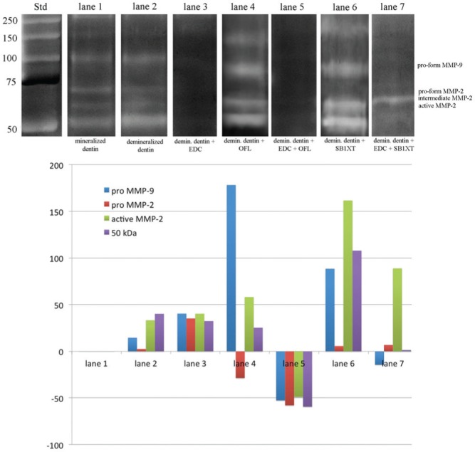
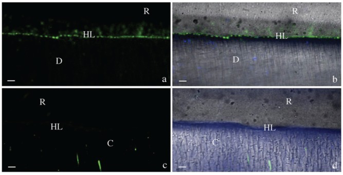
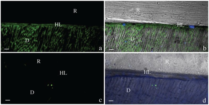
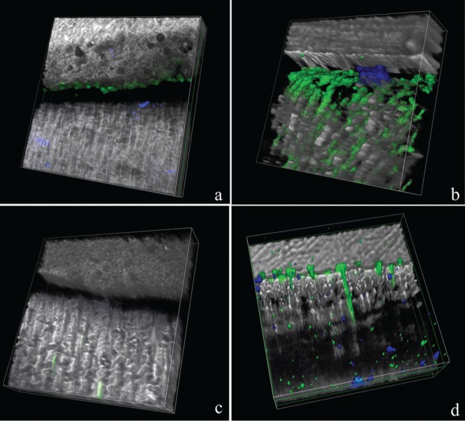
The use of protein cross-linking agents during bonding procedures has been recently proposed to improve bond durability. This study aimed to use zymography and in situ zymography techniques to evaluate the ability of 1-ethyl-3-(3-dimethylaminopropyl) carbodiimide (EDC) cross-linker to inhibit matrix metalloproteinase (MMP) activity. The hypotheses tested were that: (1) bonding procedures increase dentin gelatinolytic activity and (2) EDC pre-treatment prevents this enzymatic activity. The zymographic assay was performed on protein extracts obtained from dentin powder treated with Optibond FL or Scotchbond 1XT with or without 0.3M EDC pre-treatment. For in situ zymography, adhesive/dentin interfaces were created with the same adhesives applied to acid-etched dentin slabs pre-treated or not with EDC conditioner. Zymograms revealed increased expression of dentin endogenous MMP-2 and -9 after adhesive application, while the use of EDC as a primer inactivated dentin gelatinases. Results of in situ zymograpy showed that hybrid layers of tested adhesives exhibited intense collagenolytic activity, while almost no fluorescence signal was detected when specimens were pre-treated with EDC. The correlative analysis used in this study demonstrated that EDC could contribute to inactivate endogenous dentin MMPs within the hybrid layer created by etch-and-rinse adhesives.

Keywords: adhesive systems; biochemical assays; collagen cross-linker; dentin bonding agents; endogenous proteinases; human dentin.


Conflict of interest statement

The authors declare no potential conflicts of interest with respect to the authorship and/or publication of this article.

Figures

Figure 1.
Figure 1.
Zymographic analysis of proteins extracted from dentin powder and densitometric evaluation of bands expressed as percentage increase/decrease of MMPs activity among the different treatment groups compared with mineralized dentin (considered as baseline). Std: Standards (Std) are reported in lane Std. Lane 1: Mineralized dentin showing the presence of MMP-2 pro- and active-form (72- and 66-kDa, respectively) and pro-MMP-9 (95 kDa) and an additional band around 50 kDa. Lane 2: Proteins extracted from dentin powder demineralized with 10% phosphoric acid, showing similar presence of MMP-2 pro-form and an increase in the expression of pro-MMP-9, MMP-2 active-form, and of the additional band at 50 kDa. Lane 3: Demineralized dentin powder after incubation with 0.3M EDC showing complete inactivation of dentinal MMPs. Lane 4: Demineralized dentin powder treated with Optibond FL (OFL) showing enzymatic activation of both MMP-2 and -9 and of the additional band at approx. 50 kDa. Lane 5: Proteins extracted from demineralized dentin powder pre-treated with 0.3M EDC followed by OFL application showing complete inactivation of dentinal gelatinases. Lane 6: Demineralized dentin powder treated with Adper Scotchbond 1 XT (SB1XT) showing enzymatic activation of both MMP-2 and -9 and of the additional band at approx. 50 kDa. Lane 7: Proteins extracted from demineralized dentin powder pre-treated with 0.3M EDC followed by SB1XT application showing reduced activation of MMP-9, MMP-2, and of the 50-kDa band, and presence of the MMP-2 intermediate form compared with lane 6.
Figure 2.
Figure 2.
Resin-bonded dentin interfaces prepared with Optibond FL (OFL) with or without EDC pre-treatment, incubated with quenched fluorescein-labeled gelatin. D = Dentin; HL = Hybrid Layer; R = Resin Composite; bar = 5 µm. (a) Acquired image in green channel, showing fluorescence within the HL created with OFL. (b) Image of OFL without EDC pre-treatment, obtained by merging differential interference contrast image (showing the optical density of the resin-dentin interface) and image acquired in green channel (showing enzymatic activity). (c) Image acquired in green channel of hybrid layer created with OFL applied to acid-etched dentin pre-treated with EDC showing absence of fluorescence. (d) Image of HL created with OFL after EDC pre-treatment obtained by merging differential interference contrast image and image acquired in green channel.
Figure 3.
Figure 3.
Resin-bonded dentin interfaces prepared with Adper Scotchbond 1 XT (SB1XT) with or without EDC pre-treatment, incubated with quenched fluorescein-labeled gelatin. D = Dentin; HL = Hybrid Layer; R = Resin Composite; bar = 5 µm. (a) Image acquired in green channel, showing fluorescence (identifying intense endogenous enzymatic activity) in dentinal tubules and within the hybrid layer (HL) created with SB1XT without EDC pre-treatment. (b) Image of SB1XT without EDC pre-treatment, obtained by merging differential interference contrast image (showing the optical density of the resin-dentin interface) and image acquired in green channel (showing enzymatic activity). (c) Image acquired in green channel of hybrid layer created with SB1XT applied to acid-etched dentin pre-treated with EDC showing absence of fluorescence. (d) Image of HL created with SB1XT after EDC pre-treatment obtained by merging differential interference contrast image and image acquired in green channel.
Figure 4.
Figure 4.
Three-dimensional surface-shaded reconstruction of the acquired images. Optibond FL (OFL) and Adper Scotchbond 1 XT (SB1XT) 3D reconstructions showing intense fluorescence (evidence of gelatin hydrolysis due to endogenous proteases), throughout the hybrid layer (a and b, respectively), while reduced fluorescence was recorded when OFL and SB1XT were applied to 0.3M EDC pre-treated dentin (c and d, respectively).

References

    1. Al-Ammar A, Drummond JL, Bedran-Russo AK. (2009). The use of collagen cross-linking agents to enhance dentin bond strength. J Biomed Mater Res B Appl Biomater 91:419-424. - PMC - PubMed
    1. Almahdy A, Koller G, Sauro S, Bartsch JW, Sherriff M, Watson TF, et al. (2012). Effects of MMP inhibitors incorporated within dental adhesives. J Dent Res 91:605-611. - PMC - PubMed
    1. Atkinson SJ, Crabbe T, Cowell S, Ward RV, Butler MJ, Sato H, et al. (1995). Intermolecular autolytic cleavage can contribute to the activation of progelatinase A by cell membranes. J Biol Chem 270:30479-30485. - PubMed
    1. Avila MY, Navia JL. (2010). Effect of genipin collagen crosslinking on porcine corneas. J Cataract Refract Surg 36:659-664. - PubMed
    1. Bedran-Russo AK, Vidal CM, Dos Santos PH, Castellan CS. (2010). Long-term effect of carbodiimide on dentin matrix and resin-dentin bonds. J Biomed Mater Res B Appl Biomater 94:250-255. - PMC - PubMed

Publication types

MeSH terms

LinkOut - more resources