

Posterior fossa dural arteriovenous fistula presenting clinically as a carotid-cavernous fistula treated by a direct access cavernous sinus approach
- PMID: 24334466
- PMCID: PMC3863100
- DOI: 10.1136/bcr-2013-010939
Posterior fossa dural arteriovenous fistula presenting clinically as a carotid-cavernous fistula treated by a direct access cavernous sinus approach
Abstract
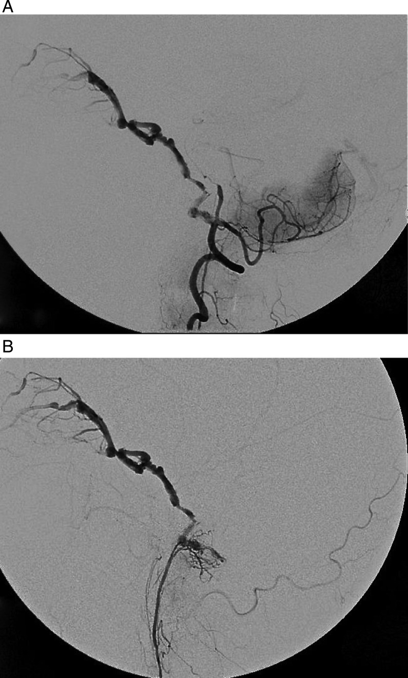
Dural arteriovenous fistulas (dAVFs) represent approximately 10-15% of all cerebral vascular malformations. Although dAVFs can occur anywhere in the brain, they occur most frequently in the cavernous and transverse-sigmoid sinuses. Posterior fossa dAVFs presenting clinically as carotid-cavernous fistulae (CCF) are rarely encountered in clinical practice. We discuss and illustrate an unusual case of a left posterior fossa dAVF that presented clinically with chemosis and early visual impairment, similar to that of CCF. This was subsequently treated by a direct access cavernous sinus approach. We describe the technique used to access the cavernous sinus directly in cases where conventional transvenous and transarterial routes have been exhausted.
Keywords: Fistula; Posterior fossa; Technique; Vascular Malformation.
Figures


Similar articles
-
Posterior fossa dural arteriovenous fistula presenting clinically as a carotid-cavernous fistula treated by a direct access cavernous sinus approach.J Neurointerv Surg. 2014 Dec;6(10):e49. doi: 10.1136/neurintsurg-2013-010939.rep. Epub 2013 Dec 18. J Neurointerv Surg. 2014. PMID: 24353329
-
[Multiple dural arteriovenous fistulas involving both the cavernous sinus and the posterior fossa: report of two cases and review of the literature].No Shinkei Geka. 2001 Nov;29(11):1065-72. No Shinkei Geka. 2001. PMID: 11758314 Review. Japanese.
-
Transorbital Cavernous Sinus Direct Puncture : Alternative to treat dural arteriovenous fistula.Clin Neuroradiol. 2018 Mar;28(1):55-61. doi: 10.1007/s00062-016-0534-z. Epub 2016 Aug 9. Clin Neuroradiol. 2018. PMID: 27506673
-
Endovascular treatment of the cavernous sinus dural arteriovenous fistula: current status and considerations.Int J Med Sci. 2020 May 1;17(8):1121-1130. doi: 10.7150/ijms.45210. eCollection 2020. Int J Med Sci. 2020. PMID: 32410842 Free PMC article. Review.
-
Development of indirect cavernous dural arteriovenous fistula after trapping for direct carotid cavernous fistula. A case report.Interv Neuroradiol. 2011 Mar;17(1):104-7. doi: 10.1177/159101991101700116. Epub 2011 Apr 29. Interv Neuroradiol. 2011. PMID: 21561566 Free PMC article.
References
-
- Kiyosue H, Hori Y, Okahara M, et al. Treatment of intracranial dural arteriovenous fistulas: current strategies based on location and hemodynamics, and alternative techniques of transcatheter embolization. Radiographics 2004;24:1637–53 - PubMed
-
- Pan HC, Sun MH, Chen WH, et al. Minimally invasive approaches to treating chemosis of the eyes from unusual dural arteriovenous fistulae. Minim Invasive Neurosurg 2009;52:222–8 - PubMed
Publication types
MeSH terms
LinkOut - more resources
Full Text Sources
Other Literature Sources