

Functional imaging biomarkers for assessing response to treatment in liver and lung metastases
- PMID: 24334562
- PMCID: PMC3864224
- DOI: 10.1102/1470-7330.2013.0047
Functional imaging biomarkers for assessing response to treatment in liver and lung metastases
Abstract
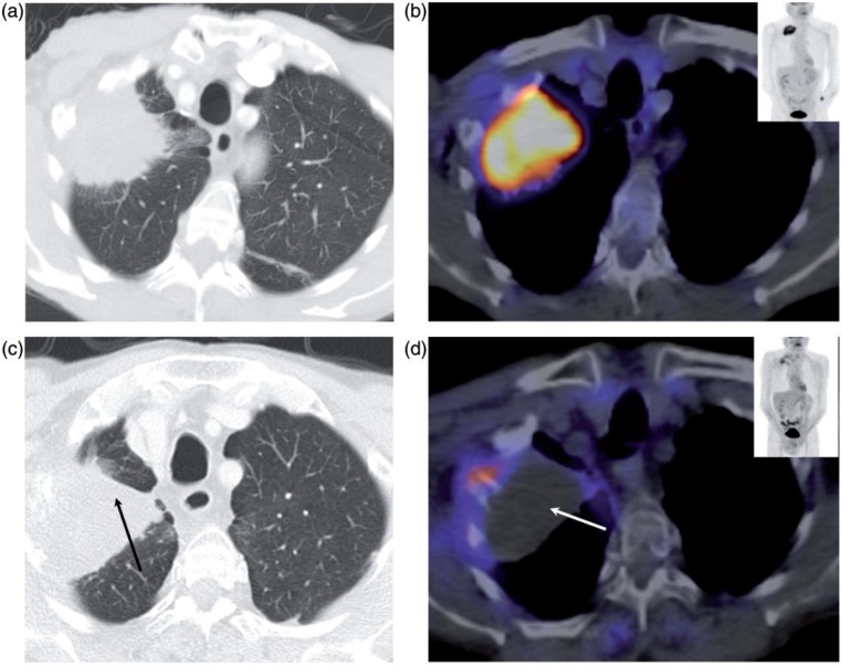
Management of patients with metastatic cancer and development of new treatments rely on imaging to provide non-invasive biomarkers of tumour response and progression. The widely used size-based criteria have increasingly become inadequate where early measures of response are required to avoid toxicity of ineffective treatments, as biological, physiologic, and molecular modifications in tumours occur before changes in gross tumour size. A multiparametric approach with the current range of imaging techniques allows functional aspects of tumours to be simultaneously interrogated. Appropriate use of these imaging techniques and their timing in relation to the treatment schedule, particularly in the context of clinical trials, is fundamental. There is a lack of consensus regarding which imaging parameters are most informative for a particular disease site and the best time to image so that, despite an increasing body of literature, open questions on these aspects remain. In addition, standardization of these new parameters is required. This review summarizes the published literature over the last decade on functional and molecular imaging techniques in assessing treatment response in liver and lung metastases.
Figures


Similar articles
-
Evaluating 90Y-glass microsphere treatment response of unresectable colorectal liver metastases by [18F]FDG PET: a comparison with CT or MRI.Eur J Nucl Med Mol Imaging. 2002 Jun;29(6):815-20. doi: 10.1007/s00259-002-0787-4. Epub 2002 Mar 29. Eur J Nucl Med Mol Imaging. 2002. PMID: 12029557 Clinical Trial.
-
PET/CT: will it change the way that we use CT in cancer imaging?Cancer Imaging. 2006 Oct 31;6(Spec No A):S52-62. doi: 10.1102/1470-7330.2006.9012. Cancer Imaging. 2006. PMID: 17114079 Free PMC article.
-
Pure ground glass nodular adenocarcinomas: Are preoperative positron emission tomography/computed tomography and brain magnetic resonance imaging useful or necessary?J Thorac Cardiovasc Surg. 2015 Sep;150(3):514-20. doi: 10.1016/j.jtcvs.2015.06.024. Epub 2015 Jun 18. J Thorac Cardiovasc Surg. 2015. PMID: 26189165
-
Respective roles of thyroglobulin, radioiodine imaging, and positron emission tomography in the assessment of thyroid cancer.Semin Nucl Med. 2006 Jul;36(3):194-205. doi: 10.1053/j.semnuclmed.2006.03.002. Semin Nucl Med. 2006. PMID: 16762610 Review.
-
Imaging modalities for the staging of patients with colorectal cancer.Neth J Med. 2012 Jan;70(1):26-34. Neth J Med. 2012. PMID: 22271811 Review.
Cited by
-
Precision of manual two-dimensional segmentations of lung and liver metastases and its impact on tumour response assessment using RECIST 1.1.Eur Radiol Exp. 2017;1(1):16. doi: 10.1186/s41747-017-0015-4. Epub 2017 Oct 30. Eur Radiol Exp. 2017. PMID: 29708185 Free PMC article.
References
-
- Harry VN, Semple SI, Parkin DE, Gilbert FJ. Use of new imaging techniques to predict tumour response to therapy. Lancet Oncol. 2010;11:92–102. - PubMed
-
- Serkova NJ, Garg K, Bradshaw-Pierce EL. Oncologic imaging end-points for the assessment of therapy response. Recent Pat Anticancer Drug Discov. 2009;4:36–53. - PubMed
-
- Sullivan DC, Gatsonis C. Response to treatment series: part 1 and introduction, measuring tumor response—challenges in the era of molecular medicine. AJR Am J Roentgenol. 2011;197:15–17. - PubMed
-
- Jaffe CC. Measures of response: RECIST, WHO, and new alternatives. J Clin Oncol. 2006;24:3245–3251. - PubMed
-
- Palmer DB. WHO handbook for reporting results of cancer treatment. Br J Cancer. 1982;45:484–485.
Publication types
MeSH terms
Substances
Grants and funding
LinkOut - more resources
Full Text Sources
Other Literature Sources
Medical