Review
. 2013 Dec 11;13(4):527-39.
doi: 10.1102/1470-7330.2013.0048.

Malignant abdominal rocks: where do they come from?

Affiliations
Review

Malignant abdominal rocks: where do they come from?

Joan M Cheng et al. Cancer Imaging. .

Abstract

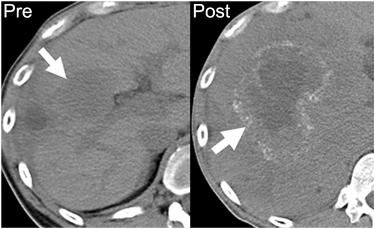
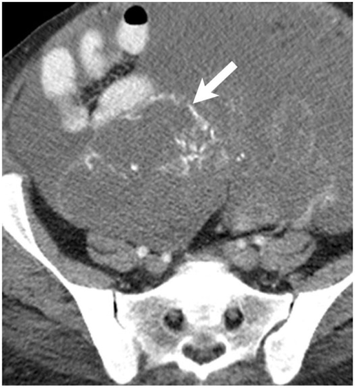
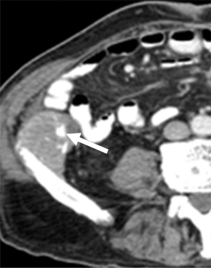
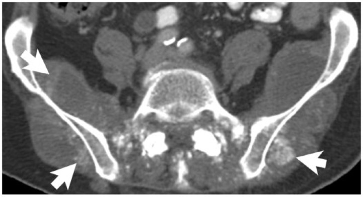
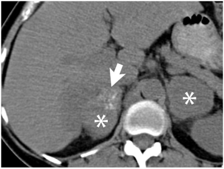
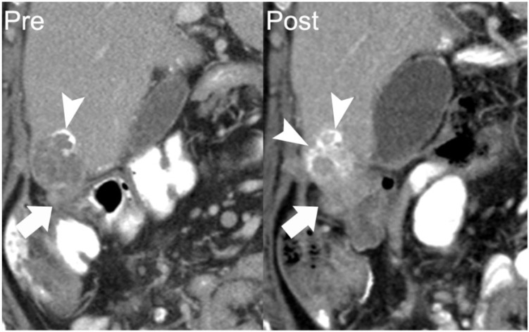
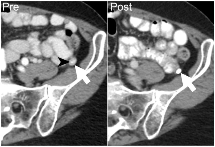
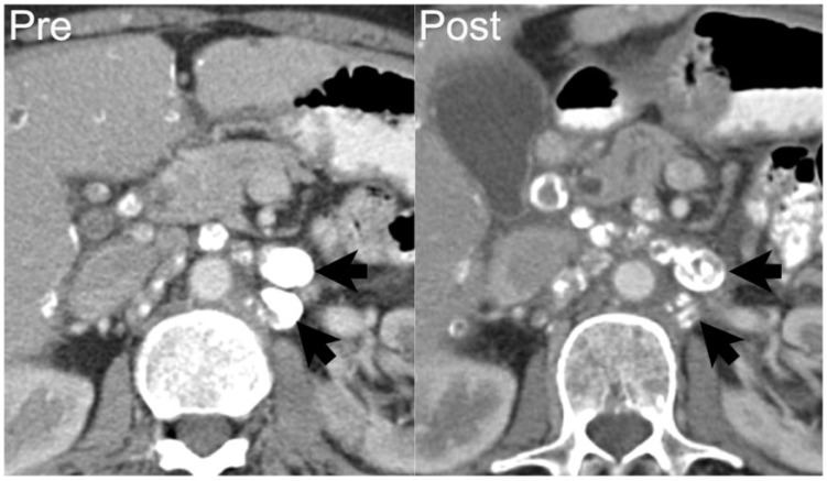
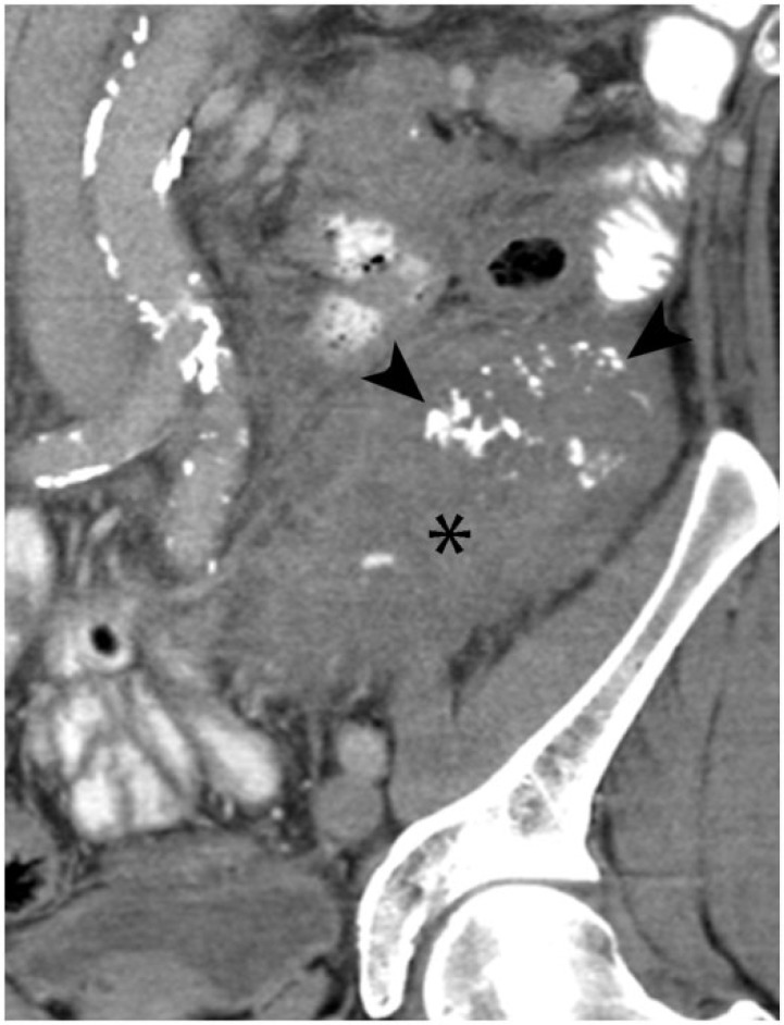
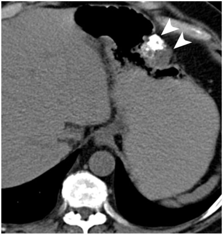
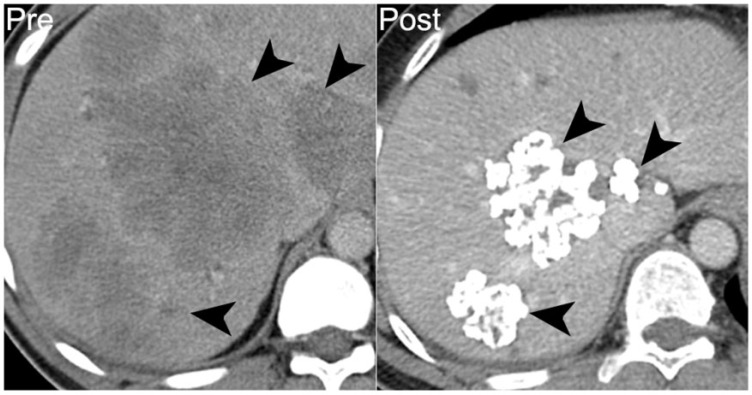
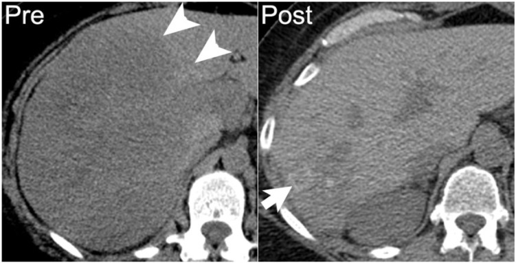
For the radiologist, calcifications in an abdominal malignancy raise questions of both diagnostic and prognostic significance. Although certain cancers are well known to calcify, such as colorectal and ovarian, malignant abdominal calcifications actually arise from a wide variety of epithelial, mesenchymal, lymphoid, or germ cell neoplasms. The pathophysiology of calcification in abdominal malignancies is heterogeneous and incompletely understood. Calcifications may present primarily, in untreated tumors, or develop during treatment; the latter can occur in variable clinical settings. A basic understanding of the varied pathogenic etiology can assist the radiologist in assessing disease status. By presenting an assortment of calcified abdominal malignancies on computed tomography in varied clinical settings, we aim not only to inform the differential diagnosis, but also to clarify the prognosis of calcifications in abdominal malignancies.


Figures

Figure 1
Figure 1
A 39-year-old man with metastatic mucinous adenocarcinoma of the distal sigmoid colon. Axial noncontrast CT image performed before chemotherapy demonstrates a noncalcified hypodense hepatic metastasis (arrow on left). After 18 months of chemotherapy on multiple regimens, there is an increase in the size of the right hepatic metastasis with a rim of amorphous calcification (arrow on right).
Figure 2
Figure 2
A 62-year-old woman with low-grade adenocarcinoma of the sigmoid colon, with focal mucinous features. Axial contrast-enhanced CT shows a curvilinear peritoneal calcification (arrow), which developed following treatment and increased with progressive disease (not shown).
Figure 3
Figure 3
A 75-year-old man with moderately differentiated mucinous adenocarcinoma of the colon. Axial contrast-enhanced CT shows a peritoneal implant with coarse peripheral calcification (arrow), which developed while on treatment and increased with progressive disease (not shown).
Figure 4
Figure 4
A 77-year-old man with rectal adenocarcinoma, without mucinous differentiation. Axial noncontrast CT image demonstrates prominent calcification (arrows) in the metastases within the gluteal and paraspinal muscles. These developed while the patient was on chemotherapy.
Figure 5
Figure 5
A 54-year-old woman with adenocarcinoma of the sigmoid colon, without mucinous differentiation. Bilateral adrenal metastases (asterisks) are seen on this axial noncontrast CT image, with amorphous calcification (arrow) within the right adrenal metastasis.
Figure 6
Figure 6
A 62-year-old woman with papillary serous ovarian adenocarcinoma with calcified peritoneal metastases. Coronal reformatted image from a contrast-enhanced CT shows a perihepatic implant with peripheral calcification (arrow on left). Follow-up CT 11 months later shows the inferomedial component of this lesion to be enlarging (arrow on right). Increasing density of calcification occurred in the setting of disease progression (arrowheads). Over this period, her cancer antigen 125 (CA125) level increased from 57 to 162 units/ml.
Figure 7
Figure 7
A 67-year-old woman with papillary serous ovarian adenocarcinoma. Axial contrast-enhanced CT performed at the start of chemotherapy shows an enhancing soft-tissue nodule in left paracolic gutter (arrow on left) with a punctate focus of calcification (arrowhead), compatible with a peritoneal implant. After 8 months of chemotherapy, follow-up contrast-enhanced CT demonstrates increased density of the calcification and decreased size of the left paracolic gutter nodule (arrow on right), indicating that increasing density of calcification may be associated with a treatment response.
Figure 8
Figure 8
A 68-year-old woman with endometrioid cystadenocarcinoma of the ovary with metastases to the retroperitoneal lymph nodes. Axial contrast-enhanced CT shows densely calcified left para-aortic lymphadenopathy (arrows on left). After 8 months of chemotherapy, disease progressed with increasing soft tissue and decreasing calcification within the nodes (arrows on right). Over this period, CA125 increased from 1439 to 3838 units/ml.
Figure 9
Figure 9
A 45-year-old man with mucinous appendiceal adenocarcinoma. Axial noncontrast CT image demonstrates scalloping (arrowheads) of the liver margin by peritoneal implants and a fine calcified septation (arrow) within the low-density ascites.
Figure 10
Figure 10
A 68-year-old man with pancreatic ductal adenocarcinoma. (A) Axial noncontrast CT image from [18F]fluorodeoxyglucose (FDG)-positron emission tomography (PET)/CT demonstrates stippled calcification in a pancreatic head mass (asterisk). (B) Fused image from FDG-PET/CT demonstrates FDG avidity engulfing the calcifications (arrows).
Figure 11
Figure 11
A 72-year-old man with pancreatic neuroendocrine tumor. Axial nonenhanced CT shows diffusely enlarged pancreatic body and tail (asterisks), with extensive calcification.
Figure 12
Figure 12
A 69-year-old man with well-differentiated neuroendocrine carcinoma of the small bowel. Coronal contrast-enhanced CT image demonstrates the typical appearance of this tumor, with a calcified mesenteric mass (arrow) and desmoplastic reaction displacing the small bowel loops peripherally.
Figure 13
Figure 13
A 55-year-old woman with medullary thyroid carcinoma. Axial noncontrast CT image demonstrates scattered extensively calcified metastases (arrowheads) and noncalcified metastases (asterisk) in the liver.
Figure 14
Figure 14
A 75-year-old man with recurrent papillary transitional cell carcinoma of the bladder. Axial nonenhanced CT image shows a metastasis in the left obturator internus, with dense calcification (arrowhead on left). Following 9 months of chemotherapy the patient’s disease progressed, with increasing size and calcification of the left obturator metastasis (arrowhead on right).
Figure 15
Figure 15
A 53-year-old woman with metastatic osteosarcoma arising from the sacrum. Mineralized lung and liver metastases are seen on axial noncontrast CT (left). She developed progressive disease after 8 months of chemotherapy, with increasing mineralization in both lung and liver metastases (right).
Figure 16
Figure 16
A 70-year-old woman with extraskeletal mesenchymal chondrosarcoma. Axial contrast-enhanced CT image demonstrates mineralized metastases in the pancreas (arrowheads) and lymph nodes (arrow). Diffuse skeletal metastases involving the vertebrae and ribs are also seen (asterisk).
Figure 17
Figure 17
A 62-year-old man with retroperitoneal dedifferentiated liposarcoma. Coronal contrast-enhanced CT image demonstrates a large left lower quadrant mass (asterisk) with coarse internal calcification (arrowhead).
Figure 18
Figure 18
A 50-year-old man with low-risk GIST. Axial noncontrast CT image performed prior to resection shows coarse calcifications in an untreated gastric GIST (arrowheads).
Figure 19
Figure 19
A 43-year-old man with high-risk jejunal GIST. Prior to treatment, axial noncontrast CT image shows diffuse hypodense hepatic metastases (arrowheads on left). After 6 years of treatment with imatinib mesylate, treated hepatic lesions are smaller and densely calcified (arrowheads on right).
Figure 20
Figure 20
A 52-year-old woman with diffuse large B-cell lymphoma. Axial noncontrast CT image demonstrates large hypodense mass in the right hepatic lobe, which was proved by biopsy to be lymphoma (arrowheads on left). One year later, following chemotherapy and stem cell transplant, the treated hepatic lesion is smaller with amorphous calcification (arrow on right).
Figure 21
Figure 21
A 40-year-old man with multiple myeloma, which had relapsed following stem cell transplant. Axial noncontrast CT image demonstrates a soft-tissue mass in the distal pancreas with peripheral calcification (asterisk). On autopsy, the pancreas was found to be entirely replaced by metastatic multiple myeloma with focal areas of necrosis.
Figure 22
Figure 22
A 45-year-old man with primary retroperitoneal malignant germ cell tumor. Axial noncontrast CT shows the untreated mass with internal calcification (arrowheads on left), which was confirmed as malignant germ cell tumor with extensive necrosis on core biopsy. Five years after neoadjuvant chemotherapy and partial resection, contrast-enhanced CT shows that the treated tumor is densely calcified with minimal visible soft-tissue component (arrowheads on right).

Similar articles

Cited by

References

    1. Porth CM. Essentials of pathophysiology: concepts of altered health states. 3rd ed. Philadelphia, PA: Lippincott Williams & Wilkins; 2010.
    1. Subhawong TK, Fishman EK, Swart JE, Carrino JA, Attar S, Fayad LM. Soft-tissue masses and mass like conditions: what does CT add to diagnosis and management? AJR Am J Roentgenol. 2010;194:1559–1567. - PMC - PubMed
    1. Siegel R, Naishadham D, Jemal A. Cancer statistics, 2013. CA-A Cancer J Clin. 2013;63:11–30. - PubMed
    1. Agarwal A, Yeh BM, Breiman BM, Qayyum A, Coakley FV. Peritoneal calcification: causes and distinguishing features on CT. Am J Roentgenol. 2004;182:441–445. - PubMed
    1. Scatarige JC, Fishman EK, Saksouk FA, Siegelman SS. Computed tomography of calcified liver masses. J Comput Assisted Tomogr. 1983;7:83–89. - PubMed

LinkOut - more resources