. 2014 May 1;23(9):2374-90.
doi: 10.1093/hmg/ddt629. Epub 2013 Dec 11.

Deletion of PDZD7 disrupts the Usher syndrome type 2 protein complex in cochlear hair cells and causes hearing loss in mice

Affiliations

Deletion of PDZD7 disrupts the Usher syndrome type 2 protein complex in cochlear hair cells and causes hearing loss in mice

Junhuang Zou et al. Hum Mol Genet. .

Abstract

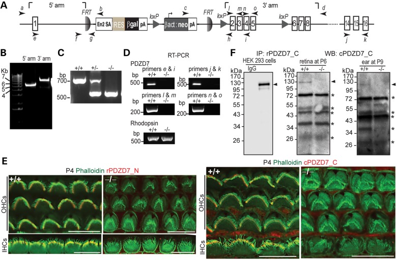
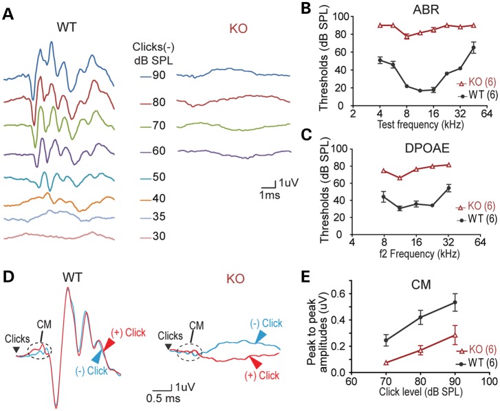
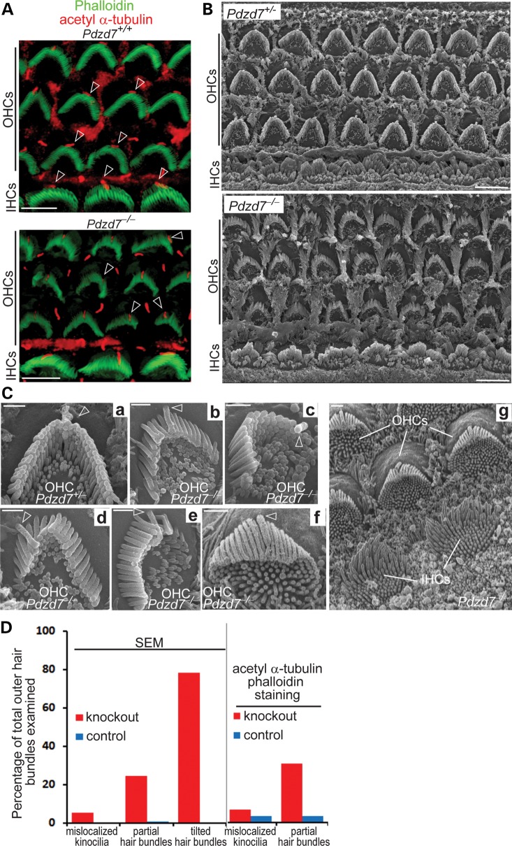
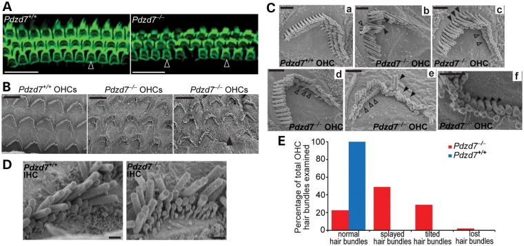
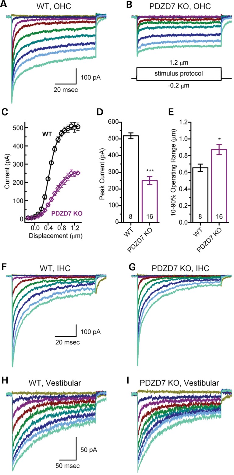
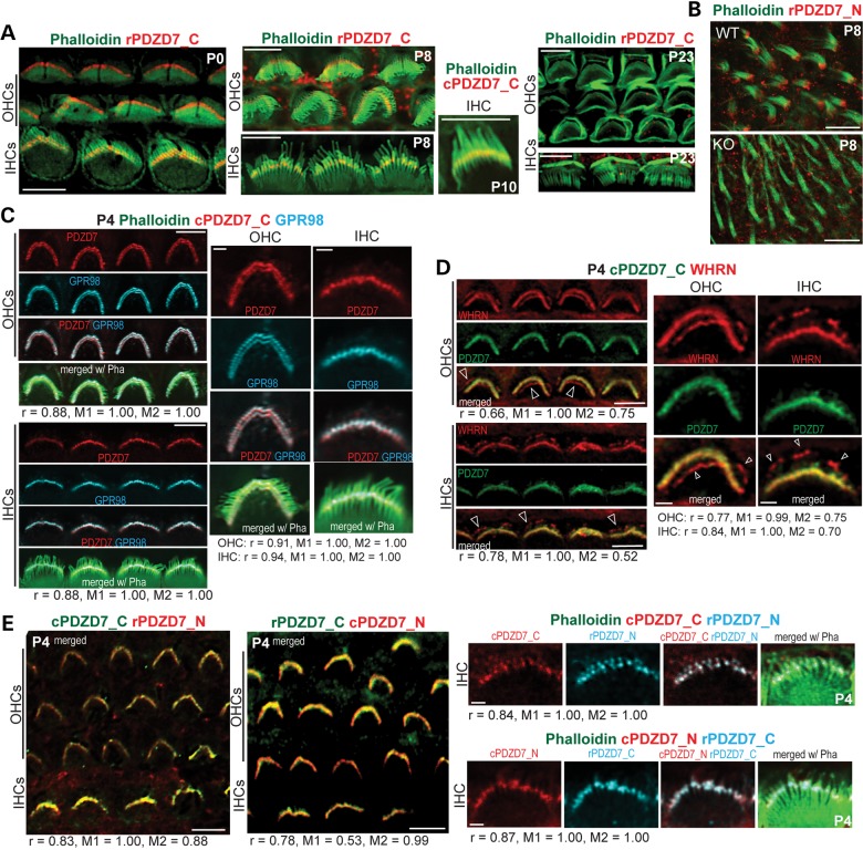
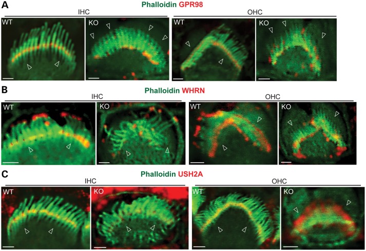
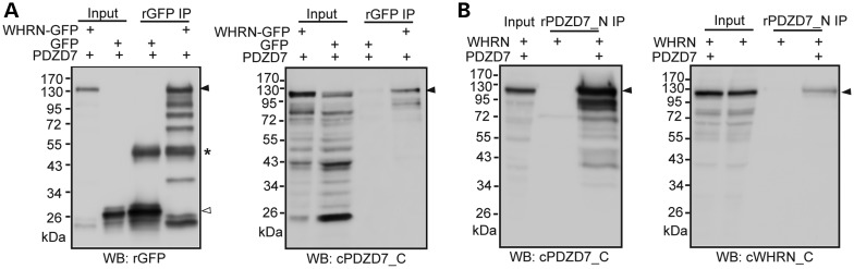
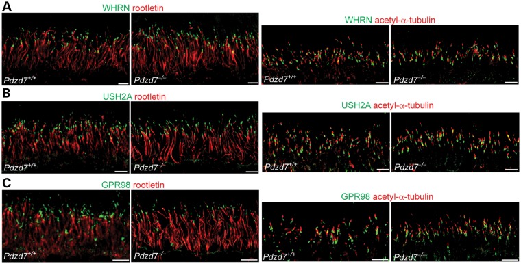
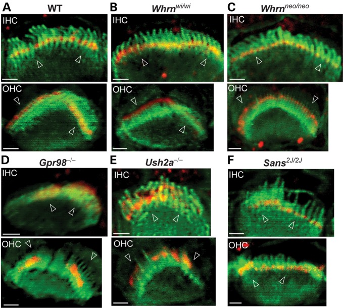
Usher syndrome type 2 (USH2) is the predominant form of USH, a leading genetic cause of combined deafness and blindness. PDZD7, a paralog of two USH causative genes, USH1C and USH2D (WHRN), was recently reported to be implicated in USH2 and non-syndromic deafness. It encodes a protein with multiple PDZ domains. To understand the biological function of PDZD7 and the pathogenic mechanism caused by PDZD7 mutations, we generated and thoroughly characterized a Pdzd7 knockout mouse model. The Pdzd7 knockout mice exhibit congenital profound deafness, as assessed by auditory brainstem response, distortion product otoacoustic emission and cochlear microphonics tests, and normal vestibular function, as assessed by their behaviors. Lack of PDZD7 leads to the disorganization of stereocilia bundles and a reduction in mechanotransduction currents and sensitivity in cochlear outer hair cells. At the molecular level, PDZD7 determines the localization of the USH2 protein complex, composed of USH2A, GPR98 and WHRN, to ankle links in developing cochlear hair cells, likely through its direct interactions with these three proteins. The localization of PDZD7 to the ankle links of cochlear hair bundles also relies on USH2 proteins. In photoreceptors of Pdzd7 knockout mice, the three USH2 proteins largely remain unchanged at the periciliary membrane complex. The electroretinogram responses of both rod and cone photoreceptors are normal in knockout mice at 1 month of age. Therefore, although the organization of the USH2 complex appears different in photoreceptors, it is clear that PDZD7 plays an essential role in organizing the USH2 complex at ankle links in developing cochlear hair cells. GenBank accession numbers: KF041446, KF041447, KF041448, KF041449, KF041450, KF041451.


Figures

Figure 1.
Figure 1.
Pdzd7−/− mice were generated. (A) A schematic diagram of the gene targeting Pdzd7tm1a(EUCOMM)Wtsi allele. Arabic numerals in rectangles denote the exon numbers. Arrows with italic lower case letters label the position of primers used in this study. En2 SA, mouse engrailed 2 gene splice acceptor; IRES, internal ribosomal entry segment; βgal, β-galactosidase gene; pA, simian virus 40 polyadenylation signal; βact, human β-actin promoter; neo, neomycin phosphotransferase gene. (B) Confirmation of insertion of the targeting cassette into the Pdzd7 gene by PCR using primer pairs of a/b and c/d. (C) Identification of the mutant allele by routine genotyping PCR using primers, f, h and g. (D) Disrupted transcription of the Pdzd7 gene shown by RT–PCR analysis in the retina using primers e, i, j, k, l, m, n and o. RT–PCR of rhodopsin was used here as a positive control. Note: primer pairs of l/m, n/o and j/k were designed to amplify PDZ1, PDZ2 and PDZ3 regions of PDZD7 cDNA, respectively. (E) Immunofluorescence demonstrates loss of PDZD7 expression in Pdzd7−/− cochlear hair cells at P4. Scale bars, 10 µm. (F) Western blotting (WB) following immunoprecipitation (IP) found loss of PDZD7 expression in the retina at P6 (middle) and in the cochlea at P9 (right) in Pdzd7−/− mice. The same procedure was performed using PDZD7-transfected HEK293 cells (left). The band just above 34 kDa on the P9 inner ear blot showed no difference between wild-type and Pdzd7−/− littermates in another experiment at P5 (data not shown). Thus, it is considered non-specific. Arrowheads point to the PDZD7 bands. Asterisks mark non-specific bands. IgG, non-immune rabbit immunoglobulin; rPDZD7_C, rabbit antibody against PDZD7_C ; cPDZD7_C, chicken antibody against PDZD7_C; rPDZD7_N, rabbit antibody against PDZD7_N. +/+, wild-type; −/−, Pdzd7 knockout. The DNA sequences of primers are shown in Supplementary material, Table S1.
Figure 2.
Figure 2.
Pdzd7−/− mice have congenital profound deafness. (A) Representative ABR responses from a pair of wild-type (WT) and Pdzd7−/− (KO) littermates at P21 in response to click stimuli. (B) ABR response thresholds at different test frequencies measured from wild-type and Pdzd7−/− littermates at P21–P28. (C) DPOAE response thresholds at different test frequencies measured from wild-type and Pdzd7−/− littermates at P21–P28. (D) Representative CMs from a pair of wild-type and Pdzd7−/− littermates at P21. (E) CM amplitudes at different click levels measured from wild-type and Pdzd7−/− littermates at P21–P28. Numbers in the parentheses are the numbers of mice tested. Error bars represent the SE.
Figure 3.
Figure 3.
Pdzd7−/− mice have disorganized outer hair bundles in the cochlea during development. (A) Staining of phalloidin (green) and acetylated α-tubulin (red) demonstrated the close to normal orientation but disrupted morphology of hair bundles (phalloidin) in the Pdzd7−/− cochlea at P4. Arrows point to kinocilia (acetylated α-tubulin) in hair bundles. Some of them moved to the side instead of being in the middle of the V-shaped hair bundles in the knockout. (B) SEM shows the morphology of hair bundles in the cochlear mid turn of Pdzd7+/− (control) and Pdzd7−/− littermates at P4. (C) High magnification view of hair bundles at P4. Compared with the Pdzd7+/− outer hair bundle (a), Pdzd7−/− outer hair bundles showed stereocilia tilting at the base toward the cell center (b, d, f and g, different regions along the cochlea), missing stereocilia (d and e) and mislocalized kinocilia (c and d). The simultaneous view of tilting OHC bundles and standing IHC bundles in Cg indicated that the tilting phenotype was not an artifact due to the imaging angle. The tilting stereocilia completely blocked the view of the two rows of shorter stereocilia. In some Pdzd7−/−OHCs, undifferentiated microvilli remained on the apex of the cells (b, c, f and g). Arrows point to kinocilia. (D) Quantification of hair bundles with mislocalized kinocilia, partial hair bundles and tilted hair bundles from SEM and fluorescence staining analyses. Details are shown in Supplementary material, Table S2. Scale bars, 5 µm (A and B) and 1 µm (C).
Figure 4.
Figure 4.
Pdzd7−/− mice show stereocilia degeneration in mature cochlear outer hair bundles. (A) The cochlear mid turns from Pdzd7+/+ and Pdzd7−/− littermates at 1 month of age were stained with phalloidin. Empty arrowheads point to the phalloidin-labeled hair bundles. (B–E) SEM analysis of hair bundles from Pdzd7+/+ and Pdzd7−/− littermates at 1 month of age. (B) Low magnification images of OHC bundles. The filled arrowhead points to a missing hair bundle. (C) Images of individual OHC bundles. Compared with the Pdzd7+/+ hair bundle (a), Pdzd7−/− hair bundles had several obvious defects: (i) the stereocilia were separated with each other (filled arrowheads in b and e); (ii) some neighboring stereocilia twisted against each other (empty arrowhead in b); (iii) the length of the stereocilia in the same row varied (filled arrowheads in c); (iv) the stereocilia in the shortest row were missing (empty arrowheads in c, d and e); (v) the stereocilia tended to lie down (filled arrowheads in e); (v) the stereocilia had different diameters in the same row (filled arrowheads in f). (D) Images of IHC bundles from Pdzd7+/+ and Pdzd7−/− littermates. (E) Quantification of defective OHC bundles from the SEM analysis. Details are shown in Supplementary material, Table S3. Scale bars, 20 µm (A), 5 µm (B) and 1 µm (C and D).
Figure 5.
Figure 5.
MET currents are abnormal in Pdzd7−/− cochlear OHCs. (A and B) Representative families of MET currents recorded from OHCs at P4 in response to mechanical hair bundle deflections that ranged from −0.2 to 1.2 µm in 0.05 µm increments. The scale bar and stimulus protocol, which shows the stimulus envelope, apply to both current families. Currents were recorded in voltage-clamp mode at −64 mV from the mid-apex. (C) Mean OHC stimulus response relations (n = 8 WT cells, 5 mice, circles; n = 16 KO cells, 7 mice, diamonds). Peak transduction currents were taken from current families and plotted as a function of stimulus amplitude. The data were shifted along the x-axis to align their mid-points, then averaged. The average data (±SE) were fitted with Boltzmann equations (lines). (D) Mean maximal MET currents (±SE) from OHCs. Genotype and number of hair cells are shown at the bottom. (E) Mean 10–90% operating range (±SE) was estimated from individual stimulus–response relations, like the one shown in (C). Genotype and number of OHCs are shown at the bottom. (F and G) Representative families of MET currents recorded from IHCs at P4 in response to mechanical hair bundle deflections. Currents were recorded in the voltage-clamp mode at −84 mV from the basal end. The scale bar applies to both panels. (H and I) Representative families of MET currents recorded from utricle type II hair cells at P4 in response to mechanical hair bundle deflections. Currents were recorded in the voltage-clamp mode at −64 mV. The scale bar applies to both panels.
Figure 6.
Figure 6.
PDZD7 is localized at ankle links in mouse inner ear hair cells. (A) Double staining of PDZD7 (red) and phalloidin (green) in cochlear hair cells. PDZD7 was localized at the base of hair bundles (phalloidin) in both IHCs and OHCs during P0 and P10. The PDZD7 signal disappeared at P23. (B) Double staining of PDZD7 (red) and phalloidin (green) in VHCs at P8. A Pdzd7−/− mouse (KO) was used as a negative control. (C) Triple staining demonstrated that PDZD7 (red) was colocalized with GPR98 (blue) at ankle links of hair bundles (phalloidin, green) ­in cochlear IHCs and OHCs at P4. Note that the four cells at the low magnification (left) and one cell at the high magnification (right) are five different cells for IHCs and OHCs. Correlation analyses between PDZD7 and GPR98 were conducted using images at both low and high magnifications. Pearson's and Manders' coefficients are shown below images. (D) Double staining demonstrated that PDZD7 (green) was colocalized with WHRN (red) at ankle links but not at the tip (arrows) of stereocilia in cochlear IHCs and OHCs at P4. Single cells in the enlarged view (right) were chosen from the four cells in the low magnification view (left) for IHCs and OHCs. Correlation analyses between PDZD7 and WHRN were conducted using images at both low and high magnifications. Pearson's and Manders' coefficients are shown below images. (E) Costaining of PDZD7_N and PDZD7_C in P4 cochlear hair cells. Left, low magnification images of both IHCs and OHCs double labeled with two combinations of PDZD7_N and PDZD7_C antibodies. Right, images of individual IHCs triple labeled with PDZD7_N, PDZD7_C and phalloidin (green). These individual cells were not chosen from the low magnification images. The colors of PDZD7_N and PDZD7_C signals match those of their word labels. Correlation analyses between signals of PDZD7_N and PDZD7_C were conducted using images at both low and high magnifications. Pearson's and Manders' coefficients are shown below images. r, Pearson's coefficient; M1, Manders' coefficient, fraction of green or blue signals overlapping red signals; M2, Manders' coefficient, fraction of red signals overlapping green or blue signals; cPDZD7_C, chicken antibody against PDZD7_C; rPDZD7_N, rabbit antibody against PDZD7_N; rPDZD7_C, rabbit antibody against PDZD7_C; cPDZD7_N, chicken antibody against PDZD7_N; Pha, phalloidin; Scale bars, 5 µm (A, low magnification images in C, D and E), 10 µm (B) and 1 µm (single cell images in C, D and E).
Figure 7.
Figure 7.
USH2 proteins are mislocalized in Pdzd7−/− cochlear hair cells. (A) GPR98 (red) was present at ankle links of hair bundles (phalloidin, green) in Pdzd7+/+ (WT) cochlear hair cells. In Pdzd7−/− mice (KO), only a small amount of GPR98 remained at ankle links. Most GPR98 moved upward toward the tip or along the stereocilia in IHC and OHC bundles. (B) WHRN (red) was present at the tip and ankle links of hair bundles (green, phalloidin) in Pdzd7+/+ cochlear hair cells. In Pdzd7−/− cochleas, the WHRN signals at the hair bundle tip stayed unchanged, while the signals at ankle links largely disappeared. (C) USH2A (red) was present at ankle links of the hair bundle (green, phalloidin) in Pdzd7+/+ cochlear hair cells. In about half of Pdzd7−/− OHCs, partial USH2A signal was mislocalized to the tip of the hair bundle, while the rest was still present at ankle links. In Pdzd7−/− IHCs, USH2A signals were significantly reduced at ankle links. Arrowheads point to the base of hair bundles. Scale bars, 1 µm.
Figure 8.
Figure 8.
PDZD7 and WHRN directly interact in HEK293 cells. (A) PDZD7 was coimmunoprecipitated with (green fluorescent protein) GFP-tagged WHRN from HEK293 cells double transfected with these two proteins. Immunoprecipitation (IP) was performed using a rabbit GFP antibody (rGFP). Cells double transfected with GFP and PDZD7 were used as a negative control. Left, the success of IP was confirmed by western blotting (WB) using the rGFP antibody. Right, the presence of PDZD7 in the immunoprecipitate was detected by western blotting using the chicken antibody against PDZD7_C (cPDZD7_C). (B) WHRN was coimmunoprecipitated with PDZD7 from HEK293 cells double transfected with these two proteins. IP was performed using the rabbit antibody against PDZD7_N. Cells transfected with only WHRN were used as a negative control. Left, the success of IP was confirmed by western blotting using the chicken antibody against PDZD7_C. Right, the presence of WHRN in the immunoprecipitate was detected by western blotting using a chicken antibody against the C-terminal region of WHRN (cWHRN_C). The filled arrowheads on the right of blots point to the targeted proteins. The empty arrowhead points to GFP bands. The asterisk labels the rabbit GFP antibody bands.
Figure 9.
Figure 9.
USH2 proteins remain at the periciliary membrane complex in Pdzd7−/− photoreceptors. Compared with wild-type (Pdzd7+/+) photoreceptors, the distributions of WHRN (green, A), USH2A (green, B) and GPR98 (green, C), relative to the ciliary rootlet (rootletin, red, left panels) and the axonemal microtubules (acetylated α-tubulin, red, right panels), appeared normal in Pdzd7−/− photoreceptors. Similar results were obtained from three pairs of Pdzd7+/+ and Pdzd7−/− mice. Scale bars, 5 µm.
Figure 10.
Figure 10.
Localization of PDZD7 relies on USH2 proteins but not SANS in cochlear hair cells. Compared with wild-type mice (WT, A), distribution of PDZD7 appeared normal or slightly diffuse at the base of the hair bundle in IHCs and OHCs of both whirler (Whrnwi/wi, B) and Whrn targeted mutant (Whrnneo/neo, C) mice. In Gpr98−/− (D) and Ush2a−/− (E) mice, PDZD7 was mislocalized along the stereocilia in both IHC and OHC bundles. In Sans2J/2J mice (F), although the hair bundle was severely disrupted, the localization of PDZD7 at ankle links was normal. Mutant mice at P4 were shown in all the panels. Red, signals from the chicken antibody against PDZD7_C; Green, hair bundles labeled with phalloidin. Arrowheads point to the base of hair bundles. Scale bars, 1 µm.

References

    1. Boughman J.A., Vernon M., Shaver K.A. Usher syndrome: definition and estimate of prevalence from two high-risk populations. J. Chronic Dis. 1983;36:595–603. - PubMed
    1. Hartong D.T., Berson E.L., Dryja T.P. Retinitis pigmentosa. Lancet. 2006;368:1795–1809. - PubMed
    1. Keats B.J., Corey D.P. The usher syndromes. Am. J. Med. Genet. 1999;89:158–166. - PubMed
    1. Eudy J.D., Weston M.D., Yao S., Hoover D.M., Rehm H.L., Ma-Edmonds M., Yan D., Ahmad I., Cheng J.J., Ayuso C., et al. Mutation of a gene encoding a protein with extracellular matrix motifs in Usher syndrome type IIa. Science. 1998;280:1753–1757. - PubMed
    1. Reiners J., Nagel-Wolfrum K., Jurgens K., Marker T., Wolfrum U. Molecular basis of human Usher syndrome: deciphering the meshes of the Usher protein network provides insights into the pathomechanisms of the Usher disease. Exp. Eye Res. 2006;83:97–119. - PubMed

Publication types

Supplementary concepts