. 2014 Jan;42(1):12-44.
doi: 10.1177/0192623313508020. Epub 2013 Dec 13.

Proceedings of the 2013 National Toxicology Program Satellite Symposium

Affiliations

Proceedings of the 2013 National Toxicology Program Satellite Symposium

Susan A Elmore et al. Toxicol Pathol. 2014 Jan.

Abstract

The 2013 annual National Toxicology Program (NTP) Satellite Symposium, entitled "Pathology Potpourri," was held in Portland, Oregon, in advance of the Society of Toxicologic Pathology's 32nd annual meeting. The goal of the NTP Symposium is to present current diagnostic pathology or nomenclature issues to the toxicologic pathology community. This article presents summaries of the speakers' presentations, including diagnostic or nomenclature issues that were presented, along with select images that were used for audience voting and discussion. Some lesions and topics covered during the symposium included a caudal tail vertebra duplication in mice; nephroblastematosis in rats; ectopic C cell tumor in a hamster; granular cell aggregates/tumor in the uterus of a hamster; Pneumocystis carinii in the lung of a rat; iatrogenic chronic inflammation in the lungs of control rats; hepatoblastoma arising within an adenoma in a mouse; humoral hypercalcemia of benignancy in a transgenic mouse; acetaminophen-induced hepatotoxicity in rats; electron microscopy images of iatrogenic intraerythrocytic inclusions in transgenic mice; questionable hepatocellular degeneration/cell death/artifact in rats; atypical endometrial hyperplasia in rats; malignant mixed Müllerian tumors/carcinosarcomas in rats; differential diagnoses of proliferative lesions of the intestine of rodents; and finally obstructive nephropathy caused by melamine poisoning in a rat.

Keywords: NTP Satellite Symposium; Pneumocystis carinii; atypical endometrial hyperplasia; bronchioloalveolar hyperplasia; duplicate vertebra; gastrointestinal diverticulum; granular cell aggregates; hepatoblastoma; hepatocellular apoptosis; intraerythrocytic inclusions; malignant mixed Müllerian tumor; nephroblastematosis; obstructive nephropathy..


Conflict of interest statement

The author(s) declared no potential conflicts of interest with respect to the research, authorship, and/or publication of this article.

Figures
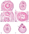
Figure 1
Figure 1
Duplicate caudal vertebra in adult B6C3F1 mice (A-F). Vehicle-control unaffected male B6C3F1 mouse (A). Transverse distal tail section exhibits single, normal distal caudal vertebra (NV) lacking processes and a vertebral arch. Note the small medullary cavity (short arrow) and normal sacrocaudal tendon/muscle bundles (long arrow). Figure B illustrates an affected male B6C3F1 mouse (low-dose group). Transverse distal tail section exhibits a supernumerary (duplicate) caudal vertebra (DV) situated dorsal to the normal vertebra (NV). Both vertebrae have small medullary cavities (arrows). The duplicate vertebra (DV) has a peripheral cartilaginous zone (CZ) and is surrounded by a fibrous capsule (FC). Figure C illustrates an affected male B6C3F1 mouse (low-dose group). Higher magnification of the tail section from (B) shows that the normal (NV) and duplicate vertebra (DV) are similarly composed of well-differentiated bone with central medullary cavities (arrows); the duplicate vertebra (DV) also has a peripheral cartilaginous zone (CZ) and is surrounded by a fibrous capsule (FC). Figure D is a higher magnification of the duplicate caudal vertebra from (B) and (C) and shows the well-differentiated bone (OS), a peripheral cartilaginous zone (CZ), and a medullary cavity (MC) containing small blood vessels and adipocytes. The duplicate vertebra is surrounded by a fibrous capsule (FC). Figure E is from an affected male B6C3F1 mouse (vehicle-control group). Transverse distal tail section exhibits a supernumerary (duplicate) caudal vertebra (DV) situated dorsal to the normal vertebra (NV). Both vertebrae lack a medullary cavity. Figure F is a transverse section of the distal tail of another affected male B6C3F1 mouse (low-dose group) and shows dorsal displacement of the supernumerary (duplicate) caudal vertebra (DV) through a dorsal skin ulceration to the external surface of the tail. The underlying normal vertebra (NV) is also displaced dorsally. Both vertebrae lack a medullary cavity. The tail subcutis (asterisk) exhibits reactive fibrosis and inflammation, and the epidermis bordering the ulcer is hyperplastic (arrow).
Figure 2
Figure 2
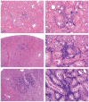
Nephroblastematosis and nephroblastoma in rat kidney. A-F, show low to high magnification views of nephroblastematosis in female Sprague Dawley rats depicting location in deep renal cortex (A,C,E), and relatively small size of focal dense interstitial accumulations of intensely staining basophilic blastema cells (B,D,F). See attempts at rosette formation in B (arrows).. Example of a differential diagnosis of focal lymphocyte accumulation (G) characterized by a population of interstitial mononuclear inflammatory cells. Well-developed nephroblastoma (H) comprised of blastema cells arranged primarily in dense mantles around ductules supported by fibrous stroma. Well-developed nephroblastoma (I) comprised of blastema cells organized into tubules that are similar to immature tubules that occur in the outer cortex of pre- and postnatal rats. The tumor is supported by a fibromyxomatous stroma. Example of a differential diagnosis of focal lymphocyte accumulation (G) characterized by a population of interstitial mononuclear inflammatory cells. Well-developed nephroblastoma (H) comprised of blastema cells arranged primarily in dense mantles around ductules supported by fibrous stroma. Well-developed nephroblastoma (I) comprised of blastema cells organized into tubules that are similar to immature tubules that occur in the outer cortex of pre- and postnatal rats. The tumor is supported by a fibromyxomatous stroma.
Figure 2
Figure 2
Nephroblastematosis and nephroblastoma in rat kidney. A-F, show low to high magnification views of nephroblastematosis in female Sprague Dawley rats depicting location in deep renal cortex (A,C,E), and relatively small size of focal dense interstitial accumulations of intensely staining basophilic blastema cells (B,D,F). See attempts at rosette formation in B (arrows).. Example of a differential diagnosis of focal lymphocyte accumulation (G) characterized by a population of interstitial mononuclear inflammatory cells. Well-developed nephroblastoma (H) comprised of blastema cells arranged primarily in dense mantles around ductules supported by fibrous stroma. Well-developed nephroblastoma (I) comprised of blastema cells organized into tubules that are similar to immature tubules that occur in the outer cortex of pre- and postnatal rats. The tumor is supported by a fibromyxomatous stroma. Example of a differential diagnosis of focal lymphocyte accumulation (G) characterized by a population of interstitial mononuclear inflammatory cells. Well-developed nephroblastoma (H) comprised of blastema cells arranged primarily in dense mantles around ductules supported by fibrous stroma. Well-developed nephroblastoma (I) comprised of blastema cells organized into tubules that are similar to immature tubules that occur in the outer cortex of pre- and postnatal rats. The tumor is supported by a fibromyxomatous stroma.
Figure 3
Figure 3
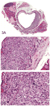
Ectopic C cell tumor in the trachea of a male Syrian (Han:AURA) hamster (A-I). A luminal cellular mass extends through the lamina propria to the level of the tracheal cartilage, adjacent to the thyroid gland (A). This mass is composed of sheets of medium-sized, polygonal to elongated cells with abundant amounts of eosinophilic, often vacuolated cytoplasm (B). The nuclei are predominantly medium-sized, round and central with coarse chromatin and occasional mitotic figures (C). Immunohistochemical stains were used to better characterize the lesion and show that the neuroendocrine cells are positive for calcitonin (D), calcitonin gene–related peptide (E), neuron-specific enolase (F), serotonin (G), and synaptophysin (H). In addition, neuroendocrine or ectopic C cells are positive with a silver stain (Grimelius) (I). A uterine granular cell lesion in a female Syrian (Han:AURA) hamster (J&K). This lesion is characterized by a large focal group of polygonal cells in the lamina propria of the uterus (J) with abundant eosinophilic granular cytoplasm and small, eccentric, nuclei with coarse chromatin (K).
Figure 3
Figure 3
Ectopic C cell tumor in the trachea of a male Syrian (Han:AURA) hamster (A-I). A luminal cellular mass extends through the lamina propria to the level of the tracheal cartilage, adjacent to the thyroid gland (A). This mass is composed of sheets of medium-sized, polygonal to elongated cells with abundant amounts of eosinophilic, often vacuolated cytoplasm (B). The nuclei are predominantly medium-sized, round and central with coarse chromatin and occasional mitotic figures (C). Immunohistochemical stains were used to better characterize the lesion and show that the neuroendocrine cells are positive for calcitonin (D), calcitonin gene–related peptide (E), neuron-specific enolase (F), serotonin (G), and synaptophysin (H). In addition, neuroendocrine or ectopic C cells are positive with a silver stain (Grimelius) (I). A uterine granular cell lesion in a female Syrian (Han:AURA) hamster (J&K). This lesion is characterized by a large focal group of polygonal cells in the lamina propria of the uterus (J) with abundant eosinophilic granular cytoplasm and small, eccentric, nuclei with coarse chromatin (K).
Figure 4
Figure 4
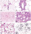
Lung lesions in F344/N control male rats from chronic oral gavage studies. For Case 1, images A and B were used to illustrate a lesion that is morphologically similar to Pneumocystis carinii infection with chronic perivascular inflammation (A) and multifocal lymphohistiocytic interstitial inflammation (B). Images C-F were presented for Case 2, which involved an oral gavage study in which corn oil served as the vehicle control. A centriacinar lesion (C) is comprised of chronic inflammation, type II epithelial hyperplasia, and bronchiolar metaplasia (D). Figure E illustrates numerous pale yellow homogeneous droplets within the alveolar spaces of a F344/N male control rat. There is positive staining of the droplets for Sudan Black indicating that the foreign material may be corn oil (F).
Figure 5
Figure 5
An hepatoblastoma arising within a pre-existing hepatic adenoma in a B6C3F1 mouse. Figure 5A shows an hepatoblastoma (arrow) associated with an expansive and well-circumscribed hepatocellular adenoma (arrowhead). Higher magnification (B) shows the two morphologically distinct neoplasms; the hepatoblastoma (arrowheads) is composed of small cells with scant cytoplasm and hyperchromatic irregular nuclei as compared to the neoplastic adenoma cells on the left. (C) The hepatoblastoma exhibits palisading of nuclei (arrowhead) and areas with widened endothelial-lined spaces containing blood (arrow) (C), and mitotic figures are frequent (arrowheads) (D).
Figure 6
Figure 6
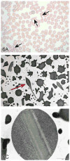
Preanalytical artifact in red blood cells in genetically engineered mice. Peripheral blood smear (A) from a genetically engineered mouse shows several abnormally shaped erythrocytes (arrows). Figures 6B and 6C are TEMs that better define the abnormally shaped erythrocytes with intracellular crystalline-like inclusions (arrows), thought to be a preanalytical artifactual change. Transmission electron microscopy (TEM) of brown fat in Wistar Han rats. Figures In these TEMs, brown fat cells characterized by fat droplets and numerous cytoplasmic mitochondria. The close proximity of the adipocytes to capillaries is illustrated in these images (arrows). Unsaturated lipid droplets appear medium to markedly electron dense (D) while saturated lipid droplets are electron lucent to flocculent or finely pale gray and granular (E).
Figure 6
Figure 6
Preanalytical artifact in red blood cells in genetically engineered mice. Peripheral blood smear (A) from a genetically engineered mouse shows several abnormally shaped erythrocytes (arrows). Figures 6B and 6C are TEMs that better define the abnormally shaped erythrocytes with intracellular crystalline-like inclusions (arrows), thought to be a preanalytical artifactual change. Transmission electron microscopy (TEM) of brown fat in Wistar Han rats. Figures In these TEMs, brown fat cells characterized by fat droplets and numerous cytoplasmic mitochondria. The close proximity of the adipocytes to capillaries is illustrated in these images (arrows). Unsaturated lipid droplets appear medium to markedly electron dense (D) while saturated lipid droplets are electron lucent to flocculent or finely pale gray and granular (E).
Figure 7
Figure 7
Small hypereosinophilic hepatocytes in female Sprague Dawley rats treated with a weak peroxisome proliferator. Figures 7A&B show medium and high magnification images of the scattered, randomly distributed, single or small clusters of affected hepatocytes (arrows). These cells are shrunken and angular with hyperchromatic nuclei and hypereosinophilic cytoplasm. Figures 7C-E are transmission electron micrographs of the affected hepatocytes. Figure 7C is an example of a hepatocyte that appears to be in an earlier stage. Note the lack of peripheralized chromatin, which would be a hallmark of early apoptosis. Also not the lack of cell swelling, which would be a hallmark of necrosis. Figure 7D is a later stage, with more condensed chromatin and cytoplasm, larger vacuoles, and adjacent/encroaching macrophages (Kupffer cells) (arrows). Figure 7E is a higher magnification of (D) showing a myelin figure that is neither within a lipid vacuole nor an autophagosome. Figure 7F is an example of classic hepatocellular apoptosis characterized by single scattered hepatocytes that are small and shrunken with hypereosinophilic cytoplasm and occasional nuclear debris. Various stages of apoptosis can be seen, including apoptotic bodies (arrows). Figure 7G is an example of classic hepatocellular necrosis, characterized by a focal group of contiguous cells with cell swelling, loss of cellular features, cell debris and inflammation. Figure 7H is an example of single cell necrosis in the liver. The affected cell (long arrow) shows marked cell swelling and karyorrhexis. The ultimate fate of such a cell is cell rupture with release of proinflammatory mediators and eventually inflammation as indicated by the short arrow.
Figure 7
Figure 7
Small hypereosinophilic hepatocytes in female Sprague Dawley rats treated with a weak peroxisome proliferator. Figures 7A&B show medium and high magnification images of the scattered, randomly distributed, single or small clusters of affected hepatocytes (arrows). These cells are shrunken and angular with hyperchromatic nuclei and hypereosinophilic cytoplasm. Figures 7C-E are transmission electron micrographs of the affected hepatocytes. Figure 7C is an example of a hepatocyte that appears to be in an earlier stage. Note the lack of peripheralized chromatin, which would be a hallmark of early apoptosis. Also not the lack of cell swelling, which would be a hallmark of necrosis. Figure 7D is a later stage, with more condensed chromatin and cytoplasm, larger vacuoles, and adjacent/encroaching macrophages (Kupffer cells) (arrows). Figure 7E is a higher magnification of (D) showing a myelin figure that is neither within a lipid vacuole nor an autophagosome. Figure 7F is an example of classic hepatocellular apoptosis characterized by single scattered hepatocytes that are small and shrunken with hypereosinophilic cytoplasm and occasional nuclear debris. Various stages of apoptosis can be seen, including apoptotic bodies (arrows). Figure 7G is an example of classic hepatocellular necrosis, characterized by a focal group of contiguous cells with cell swelling, loss of cellular features, cell debris and inflammation. Figure 7H is an example of single cell necrosis in the liver. The affected cell (long arrow) shows marked cell swelling and karyorrhexis. The ultimate fate of such a cell is cell rupture with release of proinflammatory mediators and eventually inflammation as indicated by the short arrow.
Figure 8
Figure 8
Atypical endometrial hyperplasia in the uteri of Wistar Han rats from a 2-yr toxicity/carcinogenicity bioassay. Clusters of affected glands are separated by little to no stroma (A) and lined by atypical stratified disorganized epithelium that piles up to six cell layers thick and protrudes into glandular lumens forming multiple thickened infoldings and projections (B). The epithelial cells display loss of nuclear polarization, karyomegaly, mitoses, and cellular pleomorphism (B). Also identified in this study was a papillary type of atypical hyperplasia (C) characterized by numerous small branching projections of epithelium that extend into the uterine lumen with epithelial blebbing and loss of nuclear polarization (D). Occasionally large cystic glands (E) would also show areas of similar cellular atypia (F). Focal areas of the uterine lumen (G) were also affected by atypical endometrial hyperplasia (H).
Figure 8
Figure 8
Atypical endometrial hyperplasia in the uteri of Wistar Han rats from a 2-yr toxicity/carcinogenicity bioassay. Clusters of affected glands are separated by little to no stroma (A) and lined by atypical stratified disorganized epithelium that piles up to six cell layers thick and protrudes into glandular lumens forming multiple thickened infoldings and projections (B). The epithelial cells display loss of nuclear polarization, karyomegaly, mitoses, and cellular pleomorphism (B). Also identified in this study was a papillary type of atypical hyperplasia (C) characterized by numerous small branching projections of epithelium that extend into the uterine lumen with epithelial blebbing and loss of nuclear polarization (D). Occasionally large cystic glands (E) would also show areas of similar cellular atypia (F). Focal areas of the uterine lumen (G) were also affected by atypical endometrial hyperplasia (H).
Figure 9
Figure 9
Malignant mixed Müllerian tumors (MMMTs) in female Wistar Han rats from a 2-year toxicity/carcinogenicity bioassay. The lesion presented for voting as Case 1 depicts a large solid neoplastic mass (homologous MMMT) that effaces the normal uterine architecture (A). Higher magnification of the lesion in (A) shows neoplastic cells arranged in solid sheets without the typical adenocarcinoma acinar formation (B). Neoplastic epithelium arranged in acinar structures comprises a portion of the heterologous MMMT presented in Case 2 (C). The glandular epithelium is several layers thick in some areas with very little intervening stroma between glands, characteristic of a typical uterine adenocarcinoma (C). Other portions of the mass from figure 9C are composed of small angular basophilic neoplastic cells surrounded by abundant eosinophilic material (osteoid) and islands of mature bone (D). Figures 9E&F were used to illustrate the differences in cellular morphology between a classic uterine adenocarcinoma and an MMMT. The adenocarcinoma is composed of acinar formation lined by well-differentiated neoplastic epithelium (E). The MMMT is densely cellular and solid (F). The cells contain a moderate amount of cytoplasm, indistinct cell borders, and variably sized round to oval nuclei with coarse to stippled chromatin and one to several prominent nucleoli. Cytokeratin (G) and vimentin (H) were used to illustrate the epithelial and mesenchymal components of this neoplasm, respectively.
Figure 9
Figure 9
Malignant mixed Müllerian tumors (MMMTs) in female Wistar Han rats from a 2-year toxicity/carcinogenicity bioassay. The lesion presented for voting as Case 1 depicts a large solid neoplastic mass (homologous MMMT) that effaces the normal uterine architecture (A). Higher magnification of the lesion in (A) shows neoplastic cells arranged in solid sheets without the typical adenocarcinoma acinar formation (B). Neoplastic epithelium arranged in acinar structures comprises a portion of the heterologous MMMT presented in Case 2 (C). The glandular epithelium is several layers thick in some areas with very little intervening stroma between glands, characteristic of a typical uterine adenocarcinoma (C). Other portions of the mass from figure 9C are composed of small angular basophilic neoplastic cells surrounded by abundant eosinophilic material (osteoid) and islands of mature bone (D). Figures 9E&F were used to illustrate the differences in cellular morphology between a classic uterine adenocarcinoma and an MMMT. The adenocarcinoma is composed of acinar formation lined by well-differentiated neoplastic epithelium (E). The MMMT is densely cellular and solid (F). The cells contain a moderate amount of cytoplasm, indistinct cell borders, and variably sized round to oval nuclei with coarse to stippled chromatin and one to several prominent nucleoli. Cytokeratin (G) and vimentin (H) were used to illustrate the epithelial and mesenchymal components of this neoplasm, respectively.
Figure 10
Figure 10
Three cases of gastrointestinal lesions with questions about nomenclature and differential diagnosis of both infiltration in epithelial malignancies and diverticula. Case 1 illustrates diverticula in the glandular stomach of a male CD-1 mouse characterized by cyst like structures in the tunica muscularis (A). Higher magnification (B) shows the regular differentiation of the mucosa lining the diverticulum; note also the maintained basement membrane integrity. Case 2 is illustrated by a sessile adenoma with atypical diverticula in the pyloric region of a male aryl hydrocarbon receptor knockout mouse (C) with a higher magnification (D) demonstrating the atypical character of the epithelium lining the diverticulum; note that basement membrane integrity is maintained. Case 3 was presented as an adenocarcinoma of the colon in a male F344 rat treated with once weekly subcutaneous injections of 26.6 mg/kg 1,2-dimethylhydrazine for 20 weeks; note composition of the tumor is scirrhous stroma with scattered glandular profiles covered by atypical mucosa (E). Higher magnification (F) shows areas of indistinct demarcation of the epithelial profiles from the surrounding scirrhous stroma, indicating loss of basement membrane integrity.
Figure 11
Figure 11
Foreign material in the kidney of a rat exposed to melamine and cyanuric acid orally for 12 weeks. Both the cortex and medulla show dilated renal tubules and crystalline aggregates (A). Crystalline aggregates have a characteristic “shattered plate” morphology (B). In a dilated renal tubule, foci of mineralized concretions, granular material and smaller crystals can be seen (C). Intra-tubular mineralization was often seen as concretions or concretions associated with the crystalline aggregates and (D) shows a more unusual crystalline aggregate with a structured center, possibly mineralization. Polarized light was used to show the characteristic appearance of melamine-cyanuric acid crystalline aggregates in a rat kidney (E). For comparison, polarized light was also used to show the characteristic appearance of crystalline aggregates in a dog kidney with ethylene glycol toxicity (F). Note the differences in aggregate morphology when compared to (E).

Similar articles

Cited by

References

    1. Albers TM, Simon MA, Clifford CB. Histopathology of naturally transmitted “rat respiratory virus”: progression of lesions and proposed diagnostic criteria. Vet Pathol. 2009;46:992–9. - PubMed
    1. Ameisen JC, Idziorek T, Billaut-Mulot O, Loyens M, Tissier JP, Potentier A, Ouaissi A. Apoptosis in a unicellular eukaryote (Trypanosoma cruzi): implications for the evolutionary origin and role of programmed cell death in the control of cell proliferation, differentiation and survival. Cell Death Differ. 1995;2:285–300. - PubMed
    1. Bab I, Hajbi-Yonissi C, Gabet Y, Müller R. Micro-Tomographic Atlas of the Mouse Skeleton. Springer Science + Business Media; LLC, New York, NY: 2007. pp. 87–92.
    1. Barger AM. Erythrocyte Morphology. In: Weiss Douglas J, Jane Wardrop K., editors. Schalm's Veterinary Hematology. Blackwell Publishing; Ames, IA: 2010. pp. 144–151.
    1. Barnett SA. Genotype and environment in the tail length of mice. Q J Exp Physiol. 1965;50:417–32. - PubMed

LinkOut - more resources