

Anatomic alterations in aging and age-related diseases of the eye
- PMID: 24335063
- PMCID: PMC3864374
- DOI: 10.1167/iovs.13-12711
Anatomic alterations in aging and age-related diseases of the eye
Abstract
Purpose: We described anatomic age-related changes in the human eye to determine potential areas of investigation that may lead to identifying eyes at risk for age-related disease.
Methods: A descriptive review of anatomic changes in the eye related to aging was performed in the context of current areas of investigation. The review was performed specifically for differing anatomic ocular structures, including cornea, trabecular meshwork, lens, uveal tract, Bruch's membrane, retina, RPE, vitreous, sclera, and optic nerve.
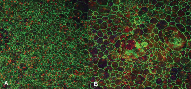
Results: Age-related changes occur in all ocular tissues. The cornea flattens and there is an attrition of endothelial cells. The shape of the trabecular meshwork changes and there is a loss of trabecular endothelium. The lens grows and becomes cataractous. The ciliary body becomes collagenized, there are choroidal vascular changes, and Bruch's membrane thickens. Retinal vessels become hyalinized and there is a loss of rods before cones in the macula. RPE morphometric changes occur with aging. The vitreous becomes liquefied and there is a loss of vitreous compartmentalization. The sclera becomes rigid and may become calcified. The optic nerve exhibits structural changes with age.
Conclusions: There are numerous anatomic age-related changes in the human eye. Current areas of investigation related to these changes include adaptive optics scanning laser ophthalmoscopy imaging of the RPE mosaic in the context of aging, and drug delivery devices that overcome age-related alterations to retinal and macular perfusion.
Keywords: aging; anatomy; pathology.
Figures
Similar articles
-
Peripapillary chorioretinal atrophy: Bruch's membrane changes and photoreceptor loss.Ophthalmology. 2000 Feb;107(2):334-43. doi: 10.1016/s0161-6420(99)00037-8. Ophthalmology. 2000. PMID: 10690836
-
Localization of myocilin/trabecular meshwork--inducible glucocorticoid response protein in the human eye.Invest Ophthalmol Vis Sci. 2000 Mar;41(3):729-40. Invest Ophthalmol Vis Sci. 2000. PMID: 10711688
-
Identification and quantitation of carotenoids and their metabolites in the tissues of the human eye.Exp Eye Res. 2001 Mar;72(3):215-23. doi: 10.1006/exer.2000.0954. Exp Eye Res. 2001. PMID: 11180970
-
[Clinical, morphological and molecular biological characteristics of the aging eye].Ophthalmologe. 2017 Feb;114(2):98-107. doi: 10.1007/s00347-016-0403-9. Ophthalmologe. 2017. PMID: 27909796 Review. German.
-
Ultrastructure of the human retina in aging and various pathological states.Micron. 2012 Jul;43(7):759-81. doi: 10.1016/j.micron.2012.01.011. Epub 2012 Feb 13. Micron. 2012. PMID: 22445096 Review.
Cited by
-
Benefits of genetic and immunohistochemical markers in understanding abnormalities in aging retina.Rom J Morphol Embryol. 2022 Jan-Mar;63(1):121-127. doi: 10.47162/RJME.63.1.12. Rom J Morphol Embryol. 2022. PMID: 36074675 Free PMC article.
-
The Role of Oxidative Stress in the Aging Eye.Life (Basel). 2023 Mar 20;13(3):837. doi: 10.3390/life13030837. Life (Basel). 2023. PMID: 36983992 Free PMC article. Review.
-
The Impact of Aging on Ocular Diseases: Unveiling Complex Interactions.Aging Dis. 2024 Sep 23;16(5):2803-2830. doi: 10.14336/AD.2024.0850. Aging Dis. 2024. PMID: 39500360 Free PMC article. Review.
-
[Forms of age-related macular degeneration].Ophthalmologe. 2015 Apr;112(4):373-85; quiz 386. doi: 10.1007/s00347-014-3186-x. Ophthalmologe. 2015. PMID: 25837316 German.
-
SOX2 haploinsufficiency promotes impaired vision at advanced age.Oncotarget. 2018 Nov 30;9(94):36684-36692. doi: 10.18632/oncotarget.26393. eCollection 2018 Nov 30. Oncotarget. 2018. PMID: 30613351 Free PMC article.
References
-
- Spencer WH. Chapter 3. Cornea. In: Spencer WH. ed Ophthalmic Pathology. An Atlas and Textbook. 4th ed. Philadelphia, PA: W. B. Saunders, Co.; 1996: 157–333
-
- Cogan DG, Kuwabara T. Arcus senilis: its pathology and histochemistry. Arch Ophthalmol. 1959; 61: 553–560 - PubMed
-
- Johnson DH, Bourne WM, Campbell RJ. The ultrastructure of Descemet's membrane. I. Change with age in normal corneas. Arch Ophthalmol. 1982; 100: 1942–1947 - PubMed
-
- Edelhauser HF. The balance between corneal transparency and edema. The Proctor lecture. Invest Ophthalmol Vis Sci. 2006; 47: 1755–1767 - PubMed
-
- Williams KK, Noe RL, Grossniklaus HE, Drews-Botsch C, Edelhauser HF. Correlation of histologic corneal endothelial cell counts with specular microscopic cell density. Arch Ophthalmol. 1992; 110: 1146–1149 - PubMed
Publication types
MeSH terms
Grants and funding
LinkOut - more resources
Full Text Sources
Other Literature Sources
Medical
