

Aging and age-related diseases of the ocular lens and vitreous body
- PMID: 24335070
- PMCID: PMC3864378
- DOI: 10.1167/iovs.13-12940
Aging and age-related diseases of the ocular lens and vitreous body
Abstract
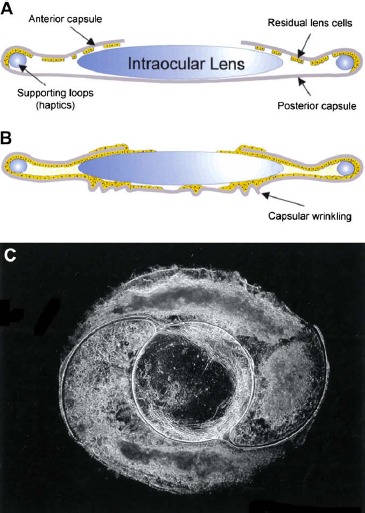
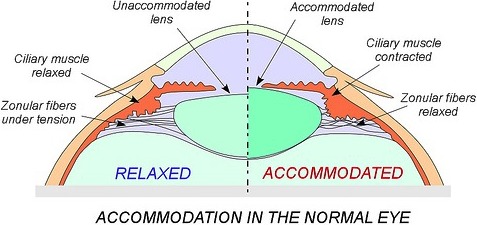
Reduced quality of life and financial burden due to visual impairment and blindness begin to increase dramatically when individuals reach the age of 40. The major causes of age-related vision loss can be traced to changes to the structure and function of the lens, one of the tissues responsible for focusing light on the retina. Age-related nuclear cataracts, which are caused by aggregation and condensation of proteins, diminish vision because they impede the transmission and focusing of light on the retina. In addition to the slow-developing age-related form, cataracts often develop rapidly as a complication of ocular surgery, such as following vitrectomy or as a consequence of vitreous gel degeneration. Posterior capsular opacification, which can develop following cataract removal, is caused by proliferation and inappropriate accumulation of lens epithelial cells on the surfaces of intraocular lenses and the posterior lens capsule. Presbyopia is a loss of accommodative amplitude and reduced ability to shift focus from far to near objects. Onset of presbyopia is associated with an increase in lens hardness and reduced ability of the lens to change shape in response to ciliary muscle contraction. Avenues of promising research that seek to delay or prevent these causes of low vision are discussed in light of our current understanding of disease pathogenesis and some challenges that must be met to achieve success.
Keywords: PCO; cataract; presbyopia; vitreous humor.
Figures
References
-
- Salm M, Belsky D, Sloan FA. Trends in cost of major eye diseases to Medicare, 1991 to 2000. Am J Ophthalmol. 2006; 142: 976–982 - PubMed
-
- Rao GN, Khanna R, Payal A. The global burden of cataract. Curr Opin Ophthalmol. 2011; 22: 4–9 - PubMed
-
- Cvekl A, Piatigorsky J. Lens development and crystallin gene expression: many roles for Pax-6. Bioessays. 1996; 18: 621–630 - PubMed
-
- Bassnett S. Lens organelle degradation. Exp Eye Res. 2002; 74: 1–6 - PubMed
-
- Nagaraj RH, Linetsky M, Stitt AW. The pathogenic role of Maillard reaction in the aging eye. Amino Acids. 2012; 42: 1205–1220 - PubMed
Publication types
MeSH terms
Grants and funding
LinkOut - more resources
Full Text Sources
Other Literature Sources
Medical
Miscellaneous
