Review
. 2013 Dec 13;54(14):ORSF81-7.
doi: 10.1167/iovs.13-12979.

Effects of diabetes on the eye

Affiliations
Review

Effects of diabetes on the eye

Gerard A Lutty. Invest Ophthalmol Vis Sci. .

Abstract

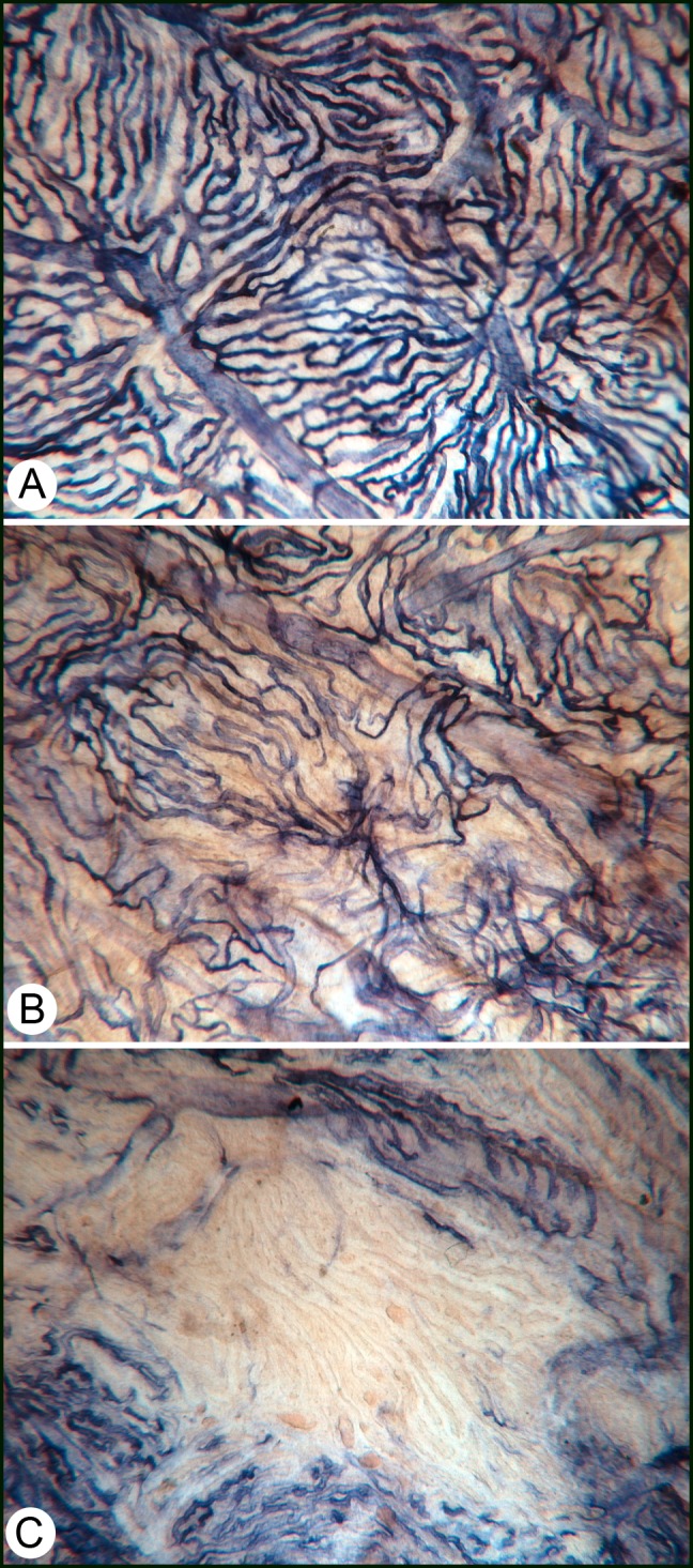
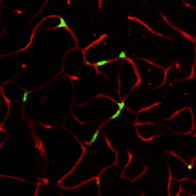
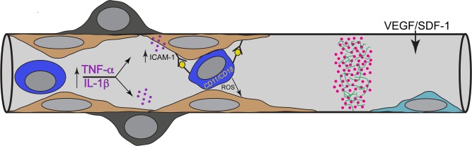
Hyperglycemia has toxic effects on almost all cells in the body. Ophthalmic complications of hyperglycemia are most profound in cornea and retina. Seventy percent of diabetics suffer from corneal complications, collectively called diabetic keratopathy, which includes include recurrent erosions, delayed wound healing, ulcers, and edema. Confocal microscopy has permitted in vivo imaging of corneal nerves, which are also affected in diabetic subjects. Gene therapies upregulating MNNG HOS transforming gene (cMet) and/or downregulating MMP10 and cathepsin S are potential future therapies for diabetic keratopathy. Diabetic retinopathy (DR) is the most common cause of blindness in people over the age of 50. There is accumulating evidence that DR is an inflammatory disease. The initial events in animal models of DR are increased vascular permeability and leukostasis. This binding of leukocytes to the endothelium results from an increase in intracellular adhesion molecule-1 (ICAM-1) on the retinal capillary endothelium (EC) and expression of CD11/CD18 on the surface of the activated leukocyte. We have observed polymorphonuclear leukocytes (PMNs) at sites of EC vascular dysfunction in diabetic retinas as well as choroid. Anti-inflammatory drugs like etanercept, aspirin, or meloxicam reduce leukostasis and EC death. Future therapies may include repopulation of the acellular capillaries after EC and pericyte death with vascular progenitors made from the patient's own blood cells.

Keywords: choroidopathy; cornea; diabetes; inflammation; retinopathy.


Figures

Figure 1
Figure 1
Adenosine diphosphatase (ADPase) activity and nonspecific esterase activity (red, indicating PMNs) in a diabetic retina (left). Bright-field (A) and dark-field illumination (B) and a section (C) after embedding in glycol methacrylate are shown for the same capillary segment (triple arrow). ADPase activity, an indicator of endothelial cell function, is lost at the site of PMN binding. When PMNs in retina were counted (D), there were significantly more PMNs in diabetic retina than in control subjects. Alkaline phosphatase activity (APase, blue) and nonspecific esterase (NSE) activity (red) in a normal choroid (E) and in a diabetic choroid (F). PMNs in the diabetic choroid are present at sites of choriocapillaris dysfunction, loss of APase activity (F). PMNs in diabetic and nondiabetic choroid were counted, and all areas of diabetic choroid had more PMNs than did normal choroid (G).
Figure 2
Figure 2
Alkaline phosphatase staining in a diabetic choroid. (A) Some areas of the choroid look normal and have organized lobules. However, on closer examination, the choriocapillaris (CC) has tortuous areas and some attenuated and constricted capillaries. (B) This area has a diffuse loss of CC, which was associated with deposits on Bruch's membrane. (C) An area of complete loss of CC, which was associated with the thickest deposits on Bruch's membrane in diabetic subjects.
Figure 3
Figure 3
Engraftment of vascular progenitors (green) into capillaries of a NOD/Scid mouse retina that experienced I/R injury. The cells were CD31+/146+ iPSCs made from cord blood CD34+ cells. The capillaries are stained with anti-collagen 4 (red).
Figure 4
Figure 4
Schematic representation of possible events in which leukostasis results in acellular capillaries. From left to right, there are high levels of TNF-α and IL-1β circulating in diabetics, which are potent upregulators of ICAM-1 by endothelial cells. ICAM-1 firmly binds neutrophils (PMNs or dark blue cell), which are activated and express CD11/CD18 on their surface. Once bound, these PMNs can create an oxidative burst, which injures endothelial cells (ECs). From repeated occlusions, ECs are lost, a platelet/fibrin thrombus (pink dots in green fibers) forms on exposed basement membrane, and eventually pericytes are lost, yielding an acellular capillary. A potential therapy might be repopulation of the acellular capillaries with vascular progenitors (light blue cell), which home to the site because of SDF-1 and VEGF upregulated expression in adjacent hypoxic retina.

Similar articles

Cited by

References

    1. Zhivov A, Winter K, Hovakimyan M, et al. Imaging and quantification of subbasal nerve plexus in healthy volunteers and diabetic patients with or without retinopathy. PLoS One. 2013; 8: e52157 - PMC - PubMed
    1. He J, Bazan HE. Mapping the nerve architecture of diabetic human corneas. Ophthalmology. 2012; 119: 956–964 - PMC - PubMed
    1. Midena E, Brugin E, Ghirlando A, Sommavilla M, Avogaro A. Corneal diabetic neuropathy: a confocal microscopy study. J Refract Surg. 2006; 22: S1047–S1052 - PubMed
    1. Midena E, Cortez M, Miotto S, Gambato C, Cavarzeran F, Ghirlando A. Confocal microscopy of corneal sub-basal nerve plexus: a quantitative and qualitative analysis in healthy and pathologic eyes. J Refract Surg. 2009; 25: S125–S130 - PubMed
    1. Chikama T, Wakuta M, Liu Y, Nishida T. Deviated mechanism of wound healing in diabetic corneas. Cornea. 2007; 26: S75–S81 - PubMed

Publication types