. 2014 Mar;42(5):3246-60.
doi: 10.1093/nar/gkt1281. Epub 2013 Dec 11.

Identification of novel methyltransferases, Bmt5 and Bmt6, responsible for the m3U methylations of 25S rRNA in Saccharomyces cerevisiae

Affiliations

Identification of novel methyltransferases, Bmt5 and Bmt6, responsible for the m3U methylations of 25S rRNA in Saccharomyces cerevisiae

Sunny Sharma et al. Nucleic Acids Res. 2014 Mar.

Abstract

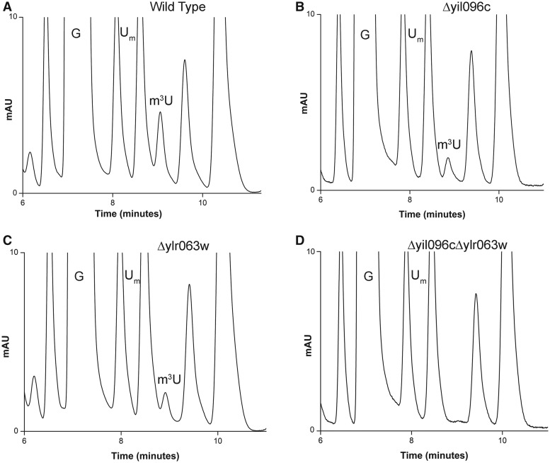
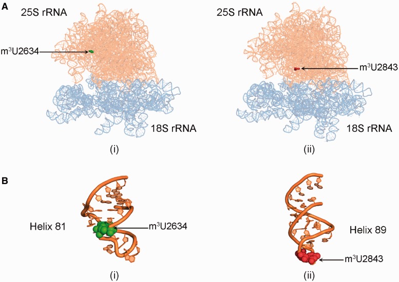
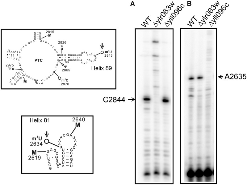
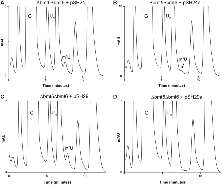
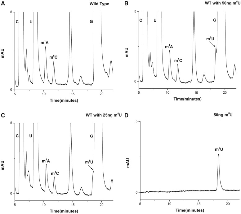
RNA contains various chemical modifications that expand its otherwise limited repertoire to mediate complex processes like translation and gene regulation. 25S rRNA of the large subunit of ribosome contains eight base methylations. Except for the methylation of uridine residues, methyltransferases for all other known base methylations have been recently identified. Here we report the identification of BMT5 (YIL096C) and BMT6 (YLR063W), two previously uncharacterized genes, to be responsible for m3U2634 and m3U2843 methylation of the 25S rRNA, respectively. These genes were identified by RP-HPLC screening of all deletion mutants of putative RNA methyltransferases and were confirmed by gene complementation and phenotypic characterization. Both proteins belong to Rossmann-fold-like methyltransferases and the point mutations in the S-adenosyl-L-methionine binding pocket abolish the methylation reaction. Bmt5 localizes in the nucleolus, whereas Bmt6 is localized predominantly in the cytoplasm. Furthermore, we showed that 25S rRNA of yeast does not contain any m5U residues as previously predicted. With Bmt5 and Bmt6, all base methyltransferases of the 25S rRNA have been identified. This will facilitate the analyses of the significance of these modifications in ribosome function and cellular physiology.


Figures

Figure 1.
Figure 1.
RP-HPLC screening of the mutants for identification of m3U methyltransferase. The 25S rRNA from mutants and isogenic wild type were digested to nucleosides using P1 nuclease and alkaline phosphatase. Nucleosides obtained after digestion was then analyzed by RP-HPLC. For optimum separation of m3U residues, we changed the elution conditions to an isocratic mode using 50% buffer A (2.5% methanol) and 50% buffer B (20% methanol). RP-HPLC chromatogram from the wild type (A), Δyil096c (B), Δylr063w (C) and Δyil096cΔylr063w (D) mutants. The peak corresponding to the m3U with a retention time of ∼9 min reduces to half in both Δyil096c and Δylr063w mutants and disappears in double mutant Δyil096cΔylr063w, highlighting the involvement of these two methyltransferases in m3U methylations of the 25S rRNA.
Figure 2.
Figure 2.
Location of m3U methylations in the ribosomal RNA. (A) 3D cartoon of the rRNA structure of ribosome. The 18S rRNA is colored light blue, whereas the 25S rRNA is shown in orange. The m3U2634 residue is displayed in green spheres (i) and the residue m3U2843 is highlighted in red spheres (ii). (B) Cartoon representing the helix 81 (i) and 85 (ii) of the 25S rRNA. The RNA is colored in orange and the modified bases U2634 and U2843 are shown as green and red spheres, respectively. The cartoon was made using PyMol software (PyMOL Molecular Graphics System, Version 1.2r3pre, Schrödinger, LLC) with PDB files, 3U5B.pdb and 3U5D.pdb.
Figure 3.
Figure 3.
Mung bean nuclease protection assay. (A) Graphic representation of Mung bean nuclease protection assay used in the present study for the analysis of specific position of modified m3U nucleoside in the Δyil096c and Δylr063w mutants. RP-HPLC chromatogram of the nucleosides derived from protected RNA fragments. Specific fragments of the 25S rRNA from wild type, Δyil096c and Δylr063w corresponding to both m3U2634 and m3U2843 were isolated. The status of m3U residue in these fragments were then analyzed by RP-HPLC. (B) RP-HPLC chromatogram of the fragment corresponding to m3U2634 isolated from the wild type and (C) from Δyil096c. (D) RP-HPLC chromatogram of the fragment corresponding to m3U2843, isolated from the wild type and (E) from Δylr063w. As evident from the chromatograms, the m3U2634 methylation is absent in Δyil096c, whereas m3U2843 is missing in Δylr063w. This clearly demonstrated that Yil096c and Ylr063w are involved in the methylation of m3U2634 and m3U2843, respectively.
Figure 4.
Figure 4.
Primer extension analysis. To further substantiate the role of Yil096c and Ylr063w in the methylation of m3U residues and validate the positions of the m3U modifications in the 25S rRNA, we performed the primer extension analysis with the 25S rRNA isolated from Δyil096c and Δylr063w deletion mutants along with isogenic wild type. The methylation of the uridine at N3 blocks the Watson–Crick base pairing, and results in a strong stop in the primer extension analysis (A) The presence of m3U at position 2843 in the 25S rRNA from the wild type and Δyil096c cells led to a strong stop at position 2844. However, this stop was absent in Δylr063w deletion mutant. (B) Similarly, owing to presence of an m3U residue at position 2634, a strong stop at position 2635 was observed in wild type and Δylr063w but was missing in Δyil096c deletion mutant. The positions of the methylated residues were determined accurately from running side by side a sequencing ladder (data not shown), is given on the sides of the gels, and the bands of interest are shown with arrows. This clearly validated the specific involvement of Ylr063w and Yil096c in the methylation of m3U2843 and m3U2634, respectively. Both helices 81 and 89, carrying m3U residues (marked with an arrow) are displayed on left of the panel A and B.
Figure 5.
Figure 5.
Gene complementation and analysis of methyltransferase dead mutants of Bmt5 and Bmt6. To confirm Bmt5 and Bmt6 to be MTases involved in performing m3U methylations, gene complementation analysis was conducted. The plasmids pSH24 and pSH29 carrying C-terminally heptahistidine-tagged Bmt5 and Bmt6, respectively, were transformed into Δbmt5Δbmt6 double mutant. The 25S rRNA from these transformed strains were then isolated and composition of 25S rRNA was analyzed with RP-HPLC. (A) Chromatogram of the 25S rRNA isolated from Δbmt5Δbmt6 strain carrying C-terminally heptahistidine-tagged Bmt5 (pSH24). (C) Chromatogram of the 25S rRNA isolated from Δbmt5Δbmt6 carrying N-terminally heptahistidine-tagged Bmt6 (pSH29). Both episomally expressed Bmt5 and Bmt6 could methylate their respective targets in vivo, seen as appearance of m3U peak in the RP-HPLC chromatogram. Furthermore, to corroborate the direct involvement of Bmt5 and Bmt6 in performing the m3U modifications, we created mutant alleles for Bmt5 and Bmt6, where we exchanged amino acids in the highly conserved SAM binding domain of both proteins. (B) RP-HPLC chromatogram from the nucleosides derived from 25S rRNA of Δbmt5Δbmt6 strain carrying mutant bmt5-G182R protein, expressed from plasmid pSH24a. (D) RP-HPLC chromatogram from the nucleosides derived from the 25S rRNA of Δbmt5Δbmt6 carrying mutant protein bmt6-G294R, expressed from plasmid pSH29a. The substitution of G182R in Bmt5 resulted in significant reduction in the amount of m3U residues, whereas the exchange of G294R in Bmt6 completely abolished the catalytic function of Bmt6.
Figure 6.
Figure 6.
Bmt5 binds SAM in vitro. To demonstrate SAM binding of Bmt5, the heterologously expressed Bmt5 was purified to homogeneity by Ni-NTA and cation exchange chromatography and its binding was analyzed by iTC. The left panel display titration of SAM into the Bmt5 protein (sample cell). The upper panel shows the baseline-corrected titration data, and the lower panel shows the binding isotherm fit to a model for a single SAM binding site. The right panel shows different parameters calculated from the titration analysis. The heat signals obtained from iTC were analyzed using Origin software supplied by Micro Cal yielding the stoichiometry (N), the equilibrium association constant Ka as well as the enthalpy (ΔH) and the entropy (ΔS) of binding. Bmt5 binds SAM with a Kd of 109 ± 10.8 µM.
Figure 7.
Figure 7.
Bmt6 interacts with Nop2 in vivo. To demonstrate the interaction of Bmt6 with the preribosomal particles and its minor nuclear localization, we immunoprecipitated the total cell extract from Bmt6-TAP–tagged strain using IgG sepharose and analyzed its interaction for Nop2 by western blotting. (A) Western blot with PAP antibodies. (B) Western blot with anti-Nop2 (EnCor Biotechnology, Florida, USA) followed by anti-mouse IgG-conjugated horseradish peroxidase (Bio-Rad; 1:10 000 dilution). The samples in the different lanes of the gels are crude extract (total cell extract), flow through (unbound cell extract) and wash fraction 6 (the beads were washed six times with IPP-150 buffer) and eluate.
Figure 8.
Figure 8.
Growth, polysome and rRNA processing analysis for Δbmt5 and Δbmt6 mutants. (A) Ten-fold serial dilutions of the strains were spotted onto solid YPD plates and were incubated at different temperatures. (B) Polysome profile of isogenic wild type (WT), Δbmt5 and Δbmt6 mutant. (C) Illustration for the 35S primary transcript. 35S rRNA contains 18, 5.8 and 25S rRNA sequences separated by ITS1 and ITS2. The processing of the 35S precursor to mature rRNA involves endonucleolytic and exonucleolytic steps at specific sites. (D) Northern blot analysis of the Δbmt5 and Δbmt6 mutant. The membrane was hybridized with radioactive (32P) labeled probes for ITS1, (f) for panel (i) and (e) for panel (ii), for ITS2, (i) for panel (iii) and to oligonucleotides specific to 18S (d) and 25S rRNA (j) for panel (iv). Loss of m3U methylations does not influence the growth and rRNA processing.
Figure 9.
Figure 9.
25S rRNA of yeast does not contain any m5U residue. (A) RP-HPLC chromatogram of 25 S rRNA displaying modified bases, m1A, m5C along with C, G and U. Surprisingly no m5U residue was detected in the chromatogram. (B) RP-HPLC chromatogram of yeast 25 S rRNA with 50 ng of m5U residues. (C) RP-HPLC chromatogram of yeast 25S rRNA with 25 ng of m5U residues. (D) RP-HPLC chromatogram of pure 50 ng m5U residues. As evident from chromatograms (B) and (C) the m5U peak could be resolved and detected in our RP-HPLC setup. This clearly demonstrates absence of m5U residues from the yeast 25S rRNA.

Similar articles

Cited by

References

    1. Ben-Shem A, Garreau de Loubresse N, Melnikov S, Jenner L, Yusupova G, Yusupov M. The structure of the eukaryotic ribosome at 3.0 A resolution. Science. 2011;334:1524–1529. - PubMed
    1. Lilley DM. The ribosome functions as a ribozyme. Chembiochem. 2001;2:31–35. - PubMed
    1. Klootwijk J, Planta RJ. Analysis of the methylation sites in yeast ribosomal RNA. Eur. J. Biochem. 1973;39:325–333. - PubMed
    1. Brand RC, Klootwijk J, Van Steenbergen TJ, De Kok AJ, Planta RJ. Secondary methylation of yeast ribosomal precursor RNA. Eur. J. Biochem. 1977;75:311–318. - PubMed
    1. Decatur WA, Fournier MJ. rRNA modifications and ribosome function. Trends Biochem. Sci. 2002;27:344–351. - PubMed

Publication types

MeSH terms